Login In

β-galactosidase-responsive Gd(Ⅲ) complexes for magnetic resonance imaging: From structural designs to diagnostic applications
- Corresponding author: Fang FANG, fang.fang@fzu.edu.cn Jianhong TANG, tangjianhong@ucas.ac.cn
Figures(7)
Citation:
Ming LIU, Xin ZHOU, Fang FANG, Yuwu ZHONG, Jianhong TANG. β-galactosidase-responsive Gd(Ⅲ) complexes for magnetic resonance imaging: From structural designs to diagnostic applications[J]. Chinese Journal of Inorganic Chemistry,
;2026, 42(11): 2275-2289.
doi:
10.11862/CJIC.20260185

Figures(7)


BRITTON M M. Magnetic resonance imaging of chemistry[J]. Chem. Soc. Rev., 2010, 39: 4036-4043
doi: 10.1039/b908397a
LI H, MEADE T J. Molecular magnetic resonance imaging with Gd(Ⅲ)-based contrast agents: Challenges and key advances[J]. J. Am. Chem. Soc., 2019, 141: 17025-17041
doi: 10.1021/jacs.9b09149
WAHSNER J, GALE E M, RODRÍGUEZ-RODRÍGUEZ A, CARAVAN P. Chemistry of MRI contrast agents: Current challenges and new frontiers[J]. Chem. Rev., 2019, 119: 957-1057
doi: 10.1021/acs.chemrev.8b00363
LAUFFER R B. Paramagnetic metal complexes as water proton relaxation agents for NMR imaging: Theory and design[J]. Chem. Rev., 1987, 87: 901-927
doi: 10.1021/cr00081a003
TERRENO E, DELLI CASTELLI D, VIALE A, AIME S. Challenges for molecular magnetic resonance imaging[J]. Chem. Rev., 2010, 110: 3019-3042
doi: 10.1021/cr100025t
KIMBERLY W T, SORBY-ADAMS A J, WEBB A G, WU E X, BEEKMAN R, BOWRY R, SCHIFF S J, DE HAVENON A, SHEN F X, SZE G, SCHAEFER P, IGLESIAS J E, ROSEN M S, SHETH K N. Brain imaging with portable low-field MRI[J]. Nat. Rev. Bioeng., 2023, 1: 617-630
doi: 10.1038/s44222-023-00086-w
ZHAO Y, DING Y, LAU V, MAN C, SU S, XIAO L, LEONG A T L, WU E X. Whole-body magnetic resonance imaging at 0.05 Tesla[J]. Science, 2024, 384: eadm7168
doi: 10.1126/science.adm7168
ALZOLA-ALDAMIZETXEBARRIA S, FERNÁNDEZ-MÉNDEZ L, PADRO D, RUÍZ-CABELLO J, RAMOS-CABRER P. A comprehensive introduction to magnetic resonance imaging relaxometry and contrast agents[J]. ACS Omega, 2022, 7: 36905-36917
doi: 10.1021/acsomega.2c03549
CARAVAN P, ELLISON J J, MCMURRY T J, LAUFFER R B. Gadolinium(Ⅲ) chelates as MRI contrast agents: Structure, dynamics, and applications[J]. Chem. Rev., 1999, 99: 2293-2352
doi: 10.1021/cr980440x
KRAS E A, SNYDER E M, SOKOLOW G E, MORROW J R. Distinct coordination chemistry of Fe(Ⅲ)-based MRI probes[J]. Accounts Chem. Res., 2022, 55: 1435-1444
doi: 10.1021/acs.accounts.2c00102
QIN R X, LI S, QIU Y W, FENG Y S, LIU Y Q, DING D D, XU L H, MA X Q, SUN W J, CHEN H M. Carbonized paramagnetic complexes of Mn(Ⅱ) as contrast agents for precise magnetic resonance imaging of sub-millimeter-sized orthotopic tumors[J]. Nat. Commun., 2022, 13: 1938
doi: 10.1038/s41467-022-29586-w
BOTÁR R, MOLNÁR E, GARDA Z, MADARASI E, TRENCSÉNYI G, KISS J, KÁLMÁN F K, TIRCSÓ G. Synthesis and characterization of a stable and inert Mn(Ⅱ)-based Zn Ⅱ responsive MRI probe for molecular imaging of glucose stimulated zinc secretion (GSZS)[J]. Inorg. Chem. Front., 2022, 9: 577-583
doi: 10.1039/D1QI00501D
AMANULLAH T A, SAMAL A K, MAYILMURUGAN R. Mn(Ⅱ) and Fe(Ⅲ) complexes as MRI contrast agents: Exploration of the second sphere relaxation for contrast enhancement[J]. Coord. Chem. Rev., 2026, 563: 217913
doi: 10.1016/j.ccr.2026.217913
LU Z R, LANEY V, LI Y. Targeted contrast agents for magnetic resonance molecular imaging of cancer[J]. Accounts Chem. Res., 2022, 55: 2833-2847
doi: 10.1021/acs.accounts.2c00346
MENG Q T, WU M M, SHANG Z Y, ZHANG Z Q, ZHANG R. Responsive gadolinium(Ⅲ) complex-based small molecule magnetic resonance imaging probes: Design, mechanism and application[J]. Coord. Chem. Rev., 2022, 457: 214398
doi: 10.1016/j.ccr.2021.214398
LI X, YUE R Y, GUAN G Q, ZHANG C, ZHOU Y, SONG G S. Recent development of pH-responsive theranostic nanoplatforms for magnetic resonance imaging-guided cancer therapy[J]. Exploration, 2023, 3: 20220002
doi: 10.1002/EXP.20220002
MORROW J R, RAYMOND J J, CHOWDHURY M S I, SAHOO P R. Redox-responsive MRI probes based on first-row transition-metal complexes[J]. Inorg. Chem., 2022, 61: 14487-14499
doi: 10.1021/acs.inorgchem.2c02197
YUE P, NAGENDRARAJ T, WANG G J, JIN Z Y, ANGELOVSKI G. The role of responsive MRI probes in the past and the future of molecular imaging[J]. Chem. Sci., 2024, 15: 20122-20154
doi: 10.1039/D4SC04849K
LIU M, GAO J P, ZHANG Y, ZHOU X, WANG Y, WU L, TIAN Z Y, TANG J H. Recent advances in bioresponsive macrocyclic gadolinium(Ⅲ) complexes for MR imaging and therapy[J]. Dalton Trans., 2025, 54: 6741-6777
doi: 10.1039/D5DT00191A
AIME S, BOTTA M, FASANO M, TERRENO E. Prototropic and water-exchange processes in aqueous solutions of Gd(Ⅲ) chelates[J]. Accounts Chem. Res., 1999, 32: 941-949
doi: 10.1021/ar970300u
TANIMOTO R, WADA T, OKADA K, SHIOMI D, SATO K, TAKUI T, SUZUKI S, NAOTA T, KOZAKI M. A molecule having 13 unpaired electrons: Magnetic property of a gadolinium(Ⅲ) complex coordinated with six nitronyl nitroxide radicals[J]. Inorg. Chem., 2022, 61: 3018-3023
doi: 10.1021/acs.inorgchem.1c03764
MCCLAIN K R, KWON H, CHAKARAWET K, NABI R, KRAGSKOW J G, CHILTON N F, BRITT R D, LONG J R, HARVEY B G. A trinuclear gadolinium cluster with a three-center one-electron bond and an S=11 ground state[J]. J. Am. Chem. Soc., 2023, 145: 8996-9002
doi: 10.1021/jacs.3c00182
BOWER D V, RICHTER J K, VON TENGG-KOBLIGK H, HEVERHAGEN J T, RUNGE V M. Gadolinium-based MRI contrast agents induce mitochondrial toxicity and cell death in human neurons, and toxicity increases with reduced kinetic stability of the agent[J]. Invest. Radiol., 2019, 54: 453-463
doi: 10.1097/RLI.0000000000000567
LIU J P, LI L, ZHANG R, XU Z P. The adjacent effect between Gd(Ⅲ) and Cu(Ⅱ) in layered double hydroxide nanoparticles synergistically enhances T1-weighted magnetic resonance imaging contrast[J]. Nanoscale Horiz., 2023, 8: 279-290
doi: 10.1039/D2NH00478J
ANTWI-BAAH R, WANG Y J, CHEN X Q, YU K. Metal-based nanoparticle magnetic resonance imaging contrast agents: Classifications, issues, and countermeasures toward their clinical translation[J]. Adv. Mater. Interfaces, 2022, 9: 2101710
doi: 10.1002/admi.202101710
UZAL-VARELA R, RODRIGUEZ-RODRIGUEZ A, WANG H, ESTEBAN-GOMEZ D, BRANDARIZ I, GALE E M, CARAVAN P, PLATAS-IGLESIAS C. Prediction of Gd(Ⅲ) complex thermodynamic stability[J]. Coord. Chem. Rev., 2022, 467: 214606
doi: 10.1016/j.ccr.2022.214606
NAVARRO R E, CORONADO A, INOUE M, OROZCO VALENCIA A N U, SOBERANES Y, SALAZAR-MEDINA A J. Gd(Ⅲ) and Yb(Ⅲ) complexes derived from a new water-soluble dioxopolyazacyclohexane macrocycle[J]. ACS Omega, 2023, 8: 34575-34582
doi: 10.1021/acsomega.3c03454
DEBRETSION A E, BUNDA S, LIHI N, PAPP G, PALLIER A, TÓTH É, KÁLMÁN F K. Exploring a rigid macrocyclic ligand bearing malonate pendants for Gd(Ⅲ) complexation: The coordination chemistry of [Gd (OPDMA)][J]. Inorg. Chem., 2025, 64: 10594-10602
doi: 10.1021/acs.inorgchem.5c01224
LUCIO-MARTíNEZ F, GARDA Z, VÁRADI B, KÁLMÁN F K, ESTEBAN-GÓMEZ D, TÓTH E V, TIRCSÓ G, PLATAS-IGLESIAS C. Rigidified derivative of the non-macrocyclic ligand H4OCTAPA for stable lanthanide(Ⅲ) complexation[J]. Inorg. Chem., 2022, 61: 5157-5171
doi: 10.1021/acs.inorgchem.2c00501
HERMANN P, KOTEK J, KUBÍČEK V, LUKEŠ I. Gadolinium(Ⅲ) complexes as MRI contrast agents: Ligand design and properties of the complexes[J]. Dalton Trans., 2008, 23: 3027-3047
PIERRE V C, BOTTA M, AIME S, RAYMOND K N. Substituent effects on Gd(Ⅲ)-based MRI contrast agents: Optimizing the stability and selectivity of the complex and the number of coordinated water molecules[J]. Inorg. Chem., 2006, 45: 8355-8364
doi: 10.1021/ic061262q
DA ROSA P P F, KITAGAWA Y, HASEGAWA Y. Luminescent lanthanide complex with seven-coordination geometry[J]. Coord. Chem. Rev., 2020, 406: 213153
doi: 10.1016/j.ccr.2019.213153
DAVIES G L, KRAMBERGER I, DAVIS J J. Environmentally responsive MRI contrast agents[J]. Chem. Commun., 2013, 49: 9704-9721
doi: 10.1039/c3cc44268c
TANG J H, LI H, YUAN C N, PARIGI G, LUCHINAT C, MEADE T J. Molecular engineering of self-immolative bioresponsive MR probes[J]. J. Am. Chem. Soc., 2023, 145: 10045-10050
doi: 10.1021/jacs.2c13672
CARRIL M. Activatable probes for diagnosis and biomarker detection by MRI[J]. J. Mater. Chem. B, 2017, 5: 4332-4347
doi: 10.1039/C7TB00093F
MALLIK R, SAHA M, SINGH V, MOHAN H, KUMARAN S S, MUKHERJEE C. Mn(Ⅱ) complex impregnated porous silica nanoparticles as Zn(Ⅱ)-responsive “Smart” MRI contrast agent for pancreas imaging[J]. J. Mater. Chem. B, 2023, 11: 8251-8261
doi: 10.1039/D3TB01289A
NING Y, ZHOU I Y, CARAVAN P. Quantitative in vivo molecular MRI[J]. Adv. Mater., 2024, 36: 2407262
doi: 10.1002/adma.202407262
WALLENFELS K, WEIL R. β-Galactosidase[M]//BOYER P D. The enzymes. California: Elsevier, 1972, 7: 617-663
KIM Y, LI H, CHOI J, BOO J, JO H, HYUN J Y, SHIN I. Glycosidase-targeting small molecules for biological and therapeutic applications[J]. Chem. Soc. Rev., 2023, 52: 7036-7070
doi: 10.1039/D3CS00032J
MOVAHEDPOUR A, AHMADI N, GHALAMFARSA F, GHESMATI Z, KHALIFEH M, MALEKSABET A, SHABANINEJAD Z, TAHERI-ANGANEH M, SAVARDASHTAKI A. β-galactosidase: From its source and applications to its recombinant form[J]. Biotechnol. Appl. Biochem., 2022, 69: 612-628
doi: 10.1002/bab.2137
KALATHINATHAN P, SAIN A, PULICHERLA K, KODIVERI MUTHUKALIANNAN G. A review on the various sources of β-galactosidase and its lactose hydrolysis property[J]. Curr. Microbiol., 2023, 80: 122
doi: 10.1007/s00284-023-03220-4
LADERO M, SANTOS A, GARCÍA-OCHOA F. Kinetic modeling of lactose hydrolysis with an immobilized β-galactosidase from Kluyveromyces fragilis[J]. Enzyme Microb. Technol., 2000, 27: 583-592
doi: 10.1016/S0141-0229(00)00244-1
SINGH P, ARORA S, RAO P S, KATHURIA D, SHARMA V, SINGH A K. Effect of process parameters on the β-galactosidase hydrolysis of lactose and galactooligosaccharide formation in concentrated skim milk[J]. Food Chem., 2022, 393: 133355
doi: 10.1016/j.foodchem.2022.133355
ZHAO J C, MU Y L, GU X Y, XU X N, GUO T T, KONG J. Site-directed mutation of β-galactosidase from Streptococcus thermophilus for galactooligosaccharide-enriched yogurt making[J]. J. Dairy Sci., 2022, 105: 940-949
doi: 10.3168/jds.2021-20905
DONG H, ZHAO L, ZHU X, WEI X H, ZHU M H, JI Q M, LUO X K, ZHANG Y T, ZHOU Y L, XU M T. Development of a novel ratiometric electrochemical sensor for monitoring β-galactosidase in Parkinson′s disease model mice[J]. Biosens. Bioelectron., 2022, 210: 114301
doi: 10.1016/j.bios.2022.114301
LI L L, JIA F F, LI Y X, PENG Y. Design strategies and biological applications of β-galactosidase fluorescent sensor in ovarian cancer research and beyond[J]. RSC Adv., 2024, 14: 3010-3023
doi: 10.1039/D3RA07968F
SU T, SHEN R J, TU D C, HAN X Y, LUO X Z, YU F B. A β-galactosidase activated near-infrared fluorescent probe for tracking cellular senescence in vitro and in vivo[J]. Smart Mol., 2025, 3: e20240062
doi: 10.1002/smo.20240062
MATSUDA J, SUZUKI O, OSHIMA A, OGURA A, NOGUCHI Y, YAMAMOTO Y, ASANO T, TAKIMOTO K, SUKEGAWA K, SUZUKI Y. β-galactosidase-deficient mouse as an animal model for GM1-gangliosidosis[J]. Glycoconjugate J., 1997, 14: 729-736
doi: 10.1023/A:1018573518127
OKADA S, O'BRIEN J S. Generalized gangliosidosis: Beta-galactosidase deficiency[J]. Science, 1968, 160: 1002-1004
doi: 10.1126/science.160.3831.1002
WU F Z, LIU J J, TAO M L, WANG M H, REN X X, HAI Z J. β-galactosidase-activatable fluorescent and photoacoustic imaging of tumor senescence[J]. Anal. Chem., 2023, 95: 10481-10485
doi: 10.1021/acs.analchem.3c01656
PAN H T, CHAI X Z, ZHANG J J. A near-infrared fluorescent probe for fast and precise imaging of senescent cells and ovarian cancer cells via tracking β-galactosidase[J]. Chin. Chem. Lett., 2023, 34: 108321
doi: 10.1016/j.cclet.2023.108321
VIJAYARAGHAVAN P, PALANISAMY S, WANG Y Y, SO P B, LIN C H, TZOU S C, YUAN S S F, WANG Y M. Senescence-associated β-galactosidase detection in human oral cancer samples using bimetallic (Fe, Cu)-MOF-919 impedimetric immunosensor[J]. Sens. Actuator Rep., 2025, 9: 100271
ROJAS-VÁZQUEZ S, LOZANO-TORRES B, GARCÍA-FERNÁNDEZ A, GALIANA I, PEREZ-VILLALBA A, MARTÍ-RODRIGO P, PALOP M J, DOMÍNGUEZ M, ORZÁEZ M, SANCENÓN F, BLANDEZ J F, FARIÑAS I, MARTÍNEZ-MÁÑEZ R. A renal clearable fluorogenic probe for in vivo β-galactosidase activity detection during aging and senolysis[J]. Nat. Commun., 2024, 15: 775
doi: 10.1038/s41467-024-44903-1
HOFER D, PAUL K, FANTUR K, BECK M, BÜRGER F, CAILLAUD C, FUMIC K, LEDVINOVA J, LUGOWSKA A, MICHELAKAKIS H, RADEVA B, RAMASWAMI U, PLECKO, B, PASCHKE E. GM1 gangliosidosis and Morquio B disease: Expression analysis of missense mutations affecting the catalytic site of acid β-galactosidase[J]. Hum. Mutat., 2009, 30: 1214-1221
doi: 10.1002/humu.21031
ALMAMMADOV T, ELMAZOGLU Z, ATAKAN G, KEPIL D, AYKENT G, KOLEMEN S, GUNBAS G. Locked and loaded: β-galactosidase activated photodynamic therapy agent enables selective imaging and targeted treatment of glioblastoma multiforme cancer cells[J]. ACS Appl. Bio Mater., 2022, 5: 4284-4293
doi: 10.1021/acsabm.2c00484
ZHANG J J, CHENG P H, PU K Y. Recent advances of molecular optical probes in imaging of β-galactosidase[J]. Bioconjug. Chem., 2019, 30: 2089-2101
doi: 10.1021/acs.bioconjchem.9b00391
YAO Y K, ZHANG Y T, YAN C X, ZHU W H, GUO Z Q. Enzyme-activatable fluorescent probes for β-galactosidase: From design to biological applications[J]. Chem. Sci., 2021, 12: 9885-9894
doi: 10.1039/D1SC02069B
CHENG S W, LIU M, WU L, TIAN Z Y, TANG J H. Non-activity-based and activatable bioresponsive peptide-conjugated Gd(Ⅲ) complexes for tumor-targeted molecular MR imaging[J]. Coord. Chem. Rev., 2026, 566: 218170
doi: 10.1016/j.ccr.2026.218170
LOUIE A Y, HÜBER M M, AHRENS E T, ROTHBÄCHER U, MOATS R, JACOBS R E, FRASER S E, MEADE T J. In vivo visualization of gene expression using magnetic resonance imaging[J]. Nat. Biotechnol., 2000, 18: 321-325
doi: 10.1038/73780
LILLEY L M, KAMPER S, CALDWELL M, CHIA Z K, BALLWEG D, VISTAIN L, KRIMMEL J, MILLS T A, MACRENARIS K, LEE P, WATERS E A, MEADE T J. Self-immolative activation of β-galactosidase-responsive probes for in vivo MR imaging in mouse models[J]. Angew. Chem. ‒Int. Edit., 2020, 59: 388-394
doi: 10.1002/anie.201909933
TANG J H, TAGHIAN T, JAYAKUMAR S, PARIGI G, LUCHINAT C, HALL E, GRAY-EDWARDS H L, MEADE T J. Bioresponsive MR imaging probes for noninvasive monitoring of AAV gene therapy[J]. Bioconjug. Chem., 2026, 37: 545-552
doi: 10.1021/acs.bioconjchem.5c00525
YU Q, ZHANG L, JIANG M, XIAO L, XIANG Y H, WANG R F, LIU Z Q, ZHOU R, YANG M H, LI C G, LIU M L, ZHOU X, CHEN S Z. An NIR fluorescence turn-on and MRl bimodal probe for concurrent real-time in vivo sensing and labeling of β-galactosidase[J]. Angew. Chem. ‒Int. Edit., 2023, 135: e202313137
doi: 10.1002/ange.202313137
LI N, XIAO L, LI S, ZHANG L, CHEN S Z. β-galactosidase-activated near-infrared fluorescence and 1H/19F MRI trimodal probe for real-time in vivo imaging of ovarian cancer[J]. Anal. Chem., 2026, 98: 5261-5270
doi: 10.1021/acs.analchem.5c05864
YU J X, KODIBAGKAR V D, HALLAC R R, LIU L, MASON R P. Dual 19F/1H MR gene reporter molecules for in vivo detection of β-galactosidase[J]. Bioconjug. Chem., 2012, 23: 596
doi: 10.1021/bc200647q
REICHERT D E, HANCOCK R D, WELCH M J. Molecular mechanics investigation of gadolinium(Ⅲ) complexes[J]. Inorg. Chem., 1996, 35: 7013-7020
doi: 10.1021/ic960495m
CHANG C A, BRITTAIN H G, TELSER J, TWEEDLE M F. pH dependence of relaxivities and hydration numbers of gadolinium(Ⅲ) complexes of linear amino carboxylates[J]. Inorg. Chem., 1990, 29: 4468-4473
doi: 10.1021/ic00347a027
PARKER D, SUTURINA E A, KUPROV I, CHILTON N F. How the ligand field in lanthanide coordination complexes determines magnetic susceptibility anisotropy, paramagnetic NMR shift, and relaxation behavior[J]. Accounts Chem. Res., 2020, 53: 1520-1534
doi: 10.1021/acs.accounts.0c00275
ROBERTSON A G, RENDINA L M. Gadolinium theranostics for the diagnosis and treatment of cancer[J]. Chem. Soc. Rev., 2021, 50: 4231-4244
doi: 10.1039/D0CS01075H
PIERRE V C, HARRIS S M, PAILLOUX S L. Comparing strategies in the design of responsive contrast agents for magnetic resonance imaging: A case study with copper and zinc[J]. Accounts Chem. Res., 2018, 51: 342-351
doi: 10.1021/acs.accounts.7b00301
YANG Y L, HU X M, YANG Z, HUANG W. Insights into molecular lanthanide complexes: Construction, properties and bioimaging and biosensing applications[J]. Adv. Funct. Mater., 2025, 35: 2412970
doi: 10.1002/adfm.202412970
BONNET C S. Zn2+ detection by MRI using Ln3+-based complexes: The central role of coordination chemistry[J]. Coord. Chem. Rev., 2018, 369: 91-104
doi: 10.1016/j.ccr.2018.04.019
PINTO S M, TOMÉ V, CALVETE M J, CASTRO M M C, TÓTH É, GERALDES C F. Metal-based redox-responsive MRI contrast agents[J]. Coord. Chem. Rev., 2019, 390: 1-31
TAGHIAN T, BATISTA A R, KAMPER S, CALDWELL M, LILLEY L, LI H, RODRIGUEZ P, MESA K, ZHENG S, KING R M, GOUNIS M J, TODEASA S, MAGUIRE A, MARTIN D R, SENA-ESTEVES M, MEADE T J, GRAY-EDWARDS H L. Real-time MR tracking of AAV gene therapy with βgal-responsive MR probe in a murine model of GM1-gangliosidosis[J]. Mol. Ther. Methods Clin. Dev., 2021, 23: 128-134
doi: 10.1016/j.omtm.2021.08.003
NERNEKLI K, MANGAROVA D B, SURYADEVARA V, HAJIPOUR M, TANG J H, WANG J, LIANG T, HARRIS M, UEYAMA T, LYONS J K, MOSELEY M E, ROUDI R, PISANI L, VON KRÜCHTEN R, DUWA R, LU-LIANG S Y, VARNIAB Z S, VASYLIV I, DAS N, MURAYAMA M, SHINOHARA I, PRATX G, GOODMAN S B, MEADE T J, DALDRUP-LINK H E. MRI detection of senescent cells in porcine knee joints with a β-galactosidase responsive Gd-chelate[J]. NPJ Imaging, 2025, 3: 18
doi: 10.1038/s44303-025-00078-y
PARK J A, LEE J J, JUNG J C, YU D Y, OH C, HA S, KIM T J, CHANG Y. Gd-DOTA conjugate of RGD as a potential tumor-targeting MRI contrast agent[J]. ChemBioChem, 2008, 9: 2811-2813
doi: 10.1002/cbic.200800529
LI W H, PARIGI G, FRAGAI M, LUCHINAT C, MEADE T J. Mechanistic studies of a calcium-dependent MRI contrast agent[J]. Inorg. Chem., 2002, 41: 4018-4024
doi: 10.1021/ic0200390
CHAUVIN T, TORRES S, ROSSETO R, KOTEK J, BADET B, DURAND P, TÓTH É. Lanthanide(Ⅲ) complexes that contain a self-immolative arm: Potential enzyme responsive contrast agents for magnetic resonance imaging[J]. Chem. ‒Eur. J., 2012, 18: 1408-1418
doi: 10.1002/chem.201101779
JOUCLAS R, LAINE S, ELISEEVA S V, MANDEL J, SZEREMETA F, RETAILLEAU P, HE J, GALLARD J F, PALLIER A, BONNET C S. Lanthanide-based probes for imaging detection of enzyme activities by NIR luminescence, T1- and ParaCEST MRI[J]. Angew. Chem. ‒Int. Edit., 2024, 63: e202317728
doi: 10.1002/anie.202317728
MOATS R A, FRASER S E, MEADE T J. A “smart” magnetic resonance imaging agent that reports on specific enzymatic activity[J]. Angew. Chem. ‒Int. Edit., 1997, 36: 726-728
doi: 10.1002/anie.199707261
URBANCZYK-PEARSON L M, FEMIA F J, SMITH J, PARIGI G, DUIMSTRA J A, ECKERMANN A L, LUCHINAT C, MEADE T J. Mechanistic investigation of β-galactosidase-activated MR contrast agents[J]. Inorg. Chem., 2008, 47: 56-68
doi: 10.1021/ic700888w
VERMA K D, MASSING J O, KAMPER S G, CARNEY C E, MACRENARIS K W, BASILION J P, MEADE T J. Synthesis and evaluation of MR probes for targeted-reporter imaging[J]. Chem. Sci., 2017, 8: 5764-5768
doi: 10.1039/C7SC02217D
AVEDANO S, BOTTA M, HAIGH J S, LONGO D L, WOODS M. Coupling fast water exchange to slow molecular tumbling in Gd3+ chelates: Why faster is not always better[J]. Inorg. Chem., 2013, 52: 8436-8450
doi: 10.1021/ic400308a
VERWILST P, PARK S, YOON B, KIM J S. Recent advances in Gd-chelate based bimodal optical/MRI contrast agents[J]. Chem. Soc. Rev., 2015, 44: 1791-1806
doi: 10.1039/C4CS00336E
CARAVAN P. Protein-targeted gadolinium-based magnetic resonance imaging (MRI) contrast agents: Design and mechanism of action[J]. Accounts Chem. Res., 2009, 42: 851-862
doi: 10.1021/ar800220p
CLOUGH T J, JIANG L, WONG K L, LONG N J. Ligand design strategies to increase stability of gadolinium-based magnetic resonance imaging contrast agents[J]. Nat. Commun., 2019, 10: 1420
doi: 10.1038/s41467-019-09342-3
SOUFI G J, HEKMATNIA A, IRAVANI S, VARMA R S. Nanoscale contrast agents for magnetic resonance imaging: A review[J]. ACS Appl. Nano Mater., 2022, 5: 10151-10166
doi: 10.1021/acsanm.2c03297
CARAVAN P. Strategies for increasing the sensitivity of gadolinium based MRI contrast agents[J]. Chem. Soc. Rev., 2006, 35: 512-523
doi: 10.1039/b510982p
DOBLE D M, BOTTA M, WANG J, AIME S, BARGE A, RAYMOND K N. Optimization of the relaxivity of MRI contrast agents: Effect of poly (ethylene glycol) chains on the water-exchange rates of GdⅢ complexes[J]. J. Am. Chem. Soc., 2001, 123: 10758-10759
doi: 10.1021/ja011085m
CHEN Y X, QIU M S, HU J S, YUE H L, YU M, CHEN S Z, ZHOU X, TAO J. Insights into host-guest interactions and enhanced MRI contrast applications of water-soluble tetrahedral metal-organic cages[J]. Sci. China‒Chem., 2025, 68: 3536-3545
WU Z Y, ZENG W W, YANG W T, YI J Y, LIU D H, XU Y, LIU C, BIAN K X, WANG H, ZHANG B B. FAP-catalyzed in situ self-assembly of magnetic resonance imaging probe for early and precise staging of liver fibrosis[J]. Sci. Adv., 2025, 11: eadt6082
doi: 10.1126/sciadv.adt6082
MAJOR J L, MEADE T J. Bioresponsive, cell-penetrating, and multimeric MR contrast agents[J]. Accounts Chem. Res., 2009, 42: 893-903
doi: 10.1021/ar800245h
CHANG Y T, CHENG C M, SU Y Z, LEE W T, HSU J S, LIU G C, CHENG T L, WANG Y M. Synthesis and characterization of a new bioactivated paramagnetic gadolinium(Ⅲ) complex [Gd (DOTA-FPG)(H2O)] for tracing gene expression[J]. Bioconjug. Chem., 2007, 18: 1716-1727
doi: 10.1021/bc070019s
HANAOKA K, KIKUCHI K, TERAI T, KOMATSU T, NAGANO T. A Gd3+-based magnetic resonance imaging contrast agent sensitive to β-galactosidase activity utilizing a receptor-induced magnetization enhancement (RIME) phenomenon[J]. Chem. ‒Eur. J., 2008, 14: 987-995
doi: 10.1002/chem.200700785
ARENA F, SINGH J B, GIANOLIO E, STEFANÌA R, AIME S. β-gal gene expression MRI reporter in melanoma tumor cells. Design, synthesis, and in vitro and in vivo testing of a Gd(Ⅲ) containing probe forming a high relaxivity, melanin-like structure upon β-gal enzymatic activation[J]. Bioconjug. Chem., 2011, 22: 2625-2635
doi: 10.1021/bc200486j
BROTHERTON A R, TSAO W, LUCHINAT C, PARIGI G, MEADE T J. Chemical control of aggregation-induced enhancement via β-galactose and gadolinium chelates[J]. Inorg. Chem., 2026, 65: 8061-8069
doi: 10.1021/acs.inorgchem.6c00654
RICCI M, RISOLO L, CARNIATO F, LALLI D, PLATAS-IGLESIAS C, BOTTA M. Field-dependent 1H relaxometry as a general probe of hydration dynamics in paramagnetic Ln3+ complexes[J]. Inorg. Chem. Front., 2026, 13: 4464-4477
doi: 10.1039/D6QI00419A
Qiuye Wang , Yabing Sun , Liangxue Lai , Haijing Cui , Yonglong Ye , Ming Yang , Weihao Zhu , Bo Yuan , Quanliang Mao , Wenzhi Ren , Aiguo Wu . MMP-9-responsive probe for fluorescence-magnetic resonance dual-mode imaging of hepatocellular carcinoma models with different metastatic capacities. Chinese Chemical Letters, 2025, 36(4): 110212-. doi: 10.1016/j.cclet.2024.110212
Meng-Juan Tang , Zhong-Hong Zhu , Hai-Ling Wang , Fu-Pei Liang , Hua-Hong Zou . Mitochondria-targeted and magnetic resonance imaging of water-stable trilobate-shaped nonanuclear lanthanide clusters. Chinese Chemical Letters, 2026, 37(9): 112126-. doi: 10.1016/j.cclet.2025.112126
Wei Zhou , Di He , Ning Liu , Ying Li , Wenzhao Han , Weiping Zhou , Siyu Zhang , Cong Yu . A PDI-based NIR-Ⅱ fluorescence imaging guided molecular phototheranostic platform for GSH-triggered gas therapy, mild photothermal therapy and NIR-activated photodynamic therapy. Chinese Chemical Letters, 2025, 36(11): 110854-. doi: 10.1016/j.cclet.2025.110854
Ling-Ling Wu , Xiangchuan Meng , Qingyang Zhang , Xiaowan Han , Feiya Yang , Qinghua Wang , Hai-Yu Hu , Nianzeng Xing . Heavy-atom engineered hypoxia-responsive probes for precisive photoacoustic imaging and cancer therapy. Chinese Chemical Letters, 2024, 35(4): 108663-. doi: 10.1016/j.cclet.2023.108663
Jinyu Guo , Yandai Lin , Shaohua He , Yueqing Chen , Fenglu Li , Renjie Ruan , Gaoxing Pan , Hexin Nan , Jibin Song , Jin Zhang . Utilizing dual-responsive iridium(Ⅲ) complex for hepatocellular carcinoma: Integrating photoacoustic imaging with chemotherapy and photodynamic therapy. Chinese Chemical Letters, 2024, 35(9): 109537-. doi: 10.1016/j.cclet.2024.109537
Mengyue Li , Yue Zheng , Zhijun Lu , Yu Yang , Jiangping Lei , Yi Teng , Wanyi Zhou , Yiman Ge , Chunhui Wu , Hong Yang , Yiyao Liu . Aptamer-targeted and pH-responsive DNA tetrahedron for survivin mRNA fluorescence imaging and chemo/gene therapy. Chinese Chemical Letters, 2026, 37(6): 111541-. doi: 10.1016/j.cclet.2025.111541
Kangren Kong , Zaiqiang Ma , Yu Yin , Zhaoming Liu , Ruikang Tang . Nuclear magnetic resonance-guided Monte Carlo/molecular dynamics structure inference for amorphous solids. Chinese Journal of Structural Chemistry, 2026, 45(3): 100773-100773. doi: 10.1016/j.cjsc.2025.100773
Siyi ZHONG , Xiaowen LIN , Jiaxin LIU , Ruyi WANG , Tao LIANG , Zhengfeng DENG , Ao ZHONG , Cuiping HAN . Targeting imaging and detection of ovarian cancer cells based on fluorescent magnetic carbon dots. Chinese Journal of Inorganic Chemistry, 2024, 40(8): 1483-1490. doi: 10.11862/CJIC.20240093
Aiai WANG , Lu ZHAO , Yunfeng BAI , Feng FENG . Research progress of bimetallic organic framework in tumor diagnosis and treatment. Chinese Journal of Inorganic Chemistry, 2024, 40(10): 1825-1839. doi: 10.11862/CJIC.20240225
Xuanxuan ZHANG , Shan YANG , Xing WANG , Fangzhu HUANG , Yujia XU , Cuiping HAN . Hyaluronic acid-modified manganese porphyrin MOF for activatable magnetic resonance imaging and synergistic sonodynamic-chemodynamic therapy performances. Chinese Journal of Inorganic Chemistry, 2026, 42(11): 2493-2503. doi: 10.11862/CJIC.20260070
Lingling Lei , Lang Liu , Yi Zhu , Yanni Wang , Zhiyao Li , Yongmin Zhang , Fengrui Yang , Weidong Pan . Molecular probes for in vivo optical imaging of chemotherapy. Chinese Chemical Letters, 2026, 37(10): 112095-. doi: 10.1016/j.cclet.2025.112095
Yiling Li , Zekun Gao , Xiuxiu Yue , Minhuan Lan , Xiuli Zheng , Benhua Wang , Shuang Zhao , Xiangzhi Song . FRET-based two-photon benzo[a] phenothiazinium photosensitizer for fluorescence imaging-guided photodynamic therapy. Chinese Chemical Letters, 2024, 35(7): 109133-. doi: 10.1016/j.cclet.2023.109133
Xu Sun , Mengchen Luo , Xinyu Xie , Mengyao Liu , Weili Wang , Tian Zhang , Lei An , Jinjun Shao , Xiaochen Dong , Yu Cai . Thienothiadiazole-based NIR-Ⅱ D-A-D dye for fluorescence and photoacoustic imaging-navigated photothermal therapy. Chinese Chemical Letters, 2026, 37(6): 111509-. doi: 10.1016/j.cclet.2025.111509
Mingrui Zhang , Lingyu Jin , Yuda Zhu , Junfeng Kou , Bo Liu , Jing Chen , Xiaolin Zhong , Xianghua Wu , Junfeng Zhang , Wenxiu Ren . A near-infrared Ⅱ fluorescent dye based on oxanthracene: Real-time imaging of drug-induced acute liver injury and photothermal therapy for tumor. Chinese Chemical Letters, 2025, 36(10): 110772-. doi: 10.1016/j.cclet.2024.110772
Ze Wang , Hao Liang , Annan Liu , Xingchen Li , Lin Guan , Lei Li , Liang He , Andrew K. Whittaker , Bai Yang , Quan Lin . Strength through unity: Alkaline phosphatase-responsive AIEgen nanoprobe for aggregation-enhanced multi-mode imaging and photothermal therapy of metastatic prostate cancer. Chinese Chemical Letters, 2025, 36(2): 109765-. doi: 10.1016/j.cclet.2024.109765
Rui WU , Yankun ZHANG , Jiufu LU , Pengfei ZHANG , Yang WANG . Research process on radioactive 18F-labelled chemical agents as positron emission tomography imaging probes for tumour detection. Chinese Journal of Inorganic Chemistry, 2025, 41(9): 1702-1718. doi: 10.11862/CJIC.20240387
Zhimin Wu , Syed Ali Abbas Abedi , Rongrong Huang , Lili Lin , Xiaogang Liu . Barrierless conical intersection as a photophysical design principle for photoacoustic and photothermal contrast agents. Chinese Chemical Letters, 2026, 37(12): 112315-. doi: 10.1016/j.cclet.2025.112315
Zhikang Wu , Guoyong Dai , Qi Li , Zheyu Wei , Shi Ru , Jianda Li , Hongli Jia , Dejin Zang , Mirjana Čolović , Yongge Wei . POV-based molecular catalysts for highly efficient esterification of alcohols with aldehydes as acylating agents. Chinese Chemical Letters, 2024, 35(8): 109061-. doi: 10.1016/j.cclet.2023.109061
Jingqi Xin , Shupeng Han , Meichen Zheng , Chenfeng Xu , Zhongxi Huang , Bin Wang , Changmin Yu , Feifei An , Yu Ren . A nitroreductase-responsive nanoprobe with homogeneous composition and high loading for preoperative non-invasive tumor imaging and intraoperative guidance. Chinese Chemical Letters, 2024, 35(7): 109165-. doi: 10.1016/j.cclet.2023.109165
Shuaige Bai , Shuai Huang , Ting Luo , Bin Feng , Yanpeng Fang , Feiyi Chu , Jie Dong , Wenbin Zeng . Debut of a responsive chemiluminescent probe for butyrylcholinesterase: Application in biological imaging and pesticide residue detection. Chinese Chemical Letters, 2025, 36(3): 110054-. doi: 10.1016/j.cclet.2024.110054
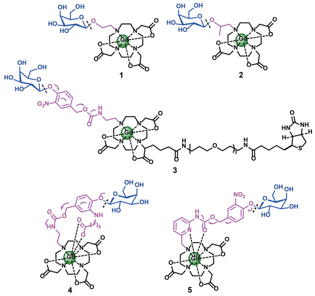
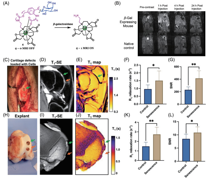
The blue color shows the β-galactosidase recognition site. The purple color indicates the bridging linker. The black color reveals the Gd(Ⅲ) MR unit. Probe 1 is the first β-galactosidase-activated Gd(Ⅲ) complex (EGad) using an ethoxy linkage to combine a galactosyl group with a DO3A-Gd MR moiety[79]. Complex 2 introduces an α-methyl group to the ethoxy linker in EGad with an enhanced MR relaxivity[59]. Probe 3 employs a carbamate--based β-galactosidase-activated self-immolative unit between a galactosyl moiety and a DO3A-Gd MR group[81]. Complex 4 attaches a pendant coordinating carboxylate ligand to the self-immolative carbamate-based chelates for in vivo detection of β-galactosidase activity[60]. Complex 5 is a pyridyl-carbamate unit based Gd(Ⅲ) with large β-galactosidase-responsive “on-off” ratio of MR relaxivity changes[34].
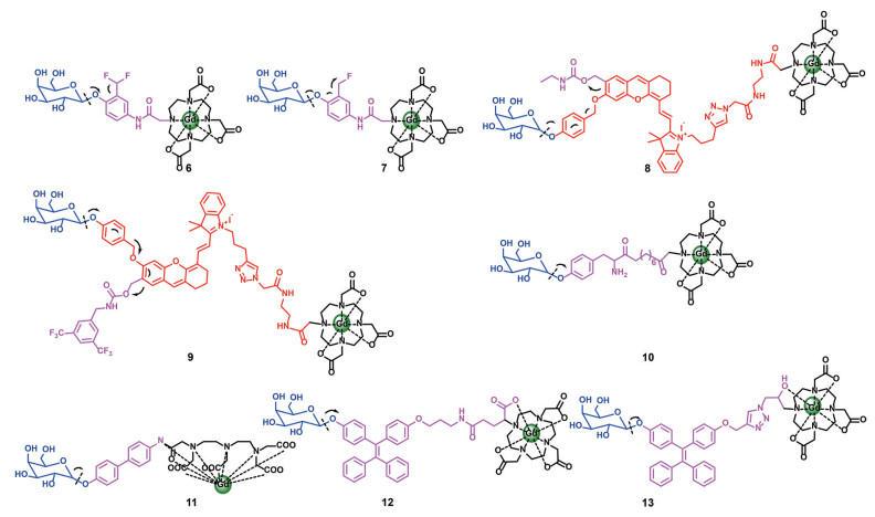
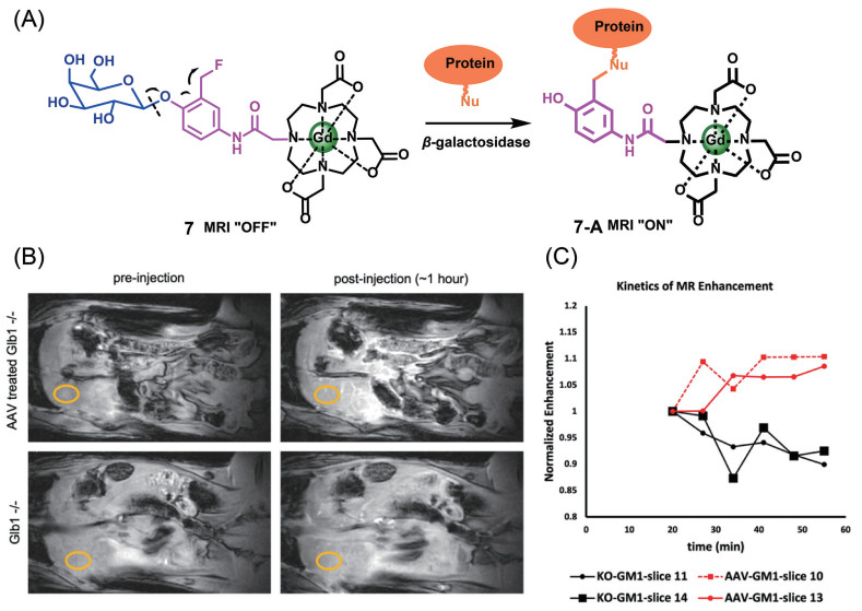
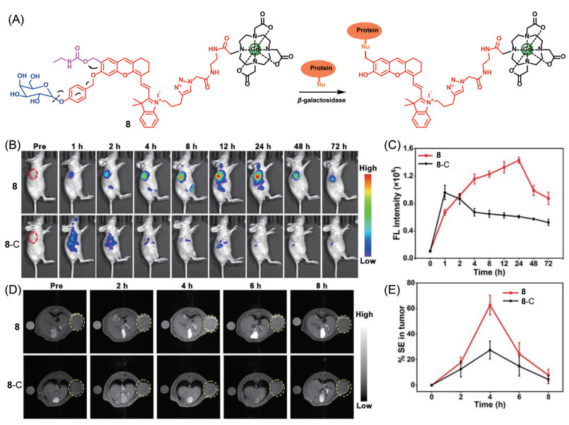
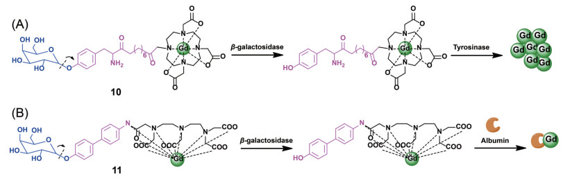
The blue color shows the β-galactosidase recognition site. The purple color indicates the bridging linker. The black color reveals the Gd(Ⅲ) MR unit. Complex 6 is a difluoromethylphenyl-β-galactopyranoside conjugated Gd(Ⅲ) agent for β-galactosidase-expressed CT26 tumor MR imaging[92]. Complex 7 contains a fluoromethyl pendant group to generate a protein-binding electrophilic quinone methide intermediate for MR imaging of AAV gene therapy[61]. Complex 8 is a β-galactosidase-responsive self-immobilizing NIR fluorescence and MR imaging bimodal agent[62]. Complex 9 represents a β-galactosidase-activated NIR fluorescence and 1H/19F MR imaging trimodal chelate for in vivo detection of ovarian cancer[63]. Probe 10 is a tyrosine-β-galactopyranoside functionalized Gd(Ⅲ), generating Gd(Ⅲ)-containing melanin oligomers or polymers upon the β-galactosidase and tyrosinase enzymatic reactions[94]. Probe 11 employs a receptor-induced magnetization enhancement (RIME) mechanism to increase protein binding affinity by changing the hydrophobicity of the agent post-enzymatic activation[93]. Complexes 12 and 13 are the β-galactopyranoside-TPE-Gd(Ⅲ) based probes to form large-sized aggregates after enzyme activation[95].
 DownLoad: