Login In
Research progress of inorganic X-ray nanoscintillators
- Corresponding author: Yejun ZHANG, yjzhang2009@sinano.ac.cn Yu ZOU, yzou2012@sinano.ac.cn Qiangbin WANG, qbwang2008@sinano.ac.cn
Figures(13)
Citation:
Xiaoyu YANG, Yejun ZHANG, Yu ZOU, Hongchao YANG, Jiang JIANG, Qiangbin WANG. Research progress of inorganic X-ray nanoscintillators[J]. Chinese Journal of Inorganic Chemistry,
;2025, 41(10): 1929-1952.
doi:
10.11862/CJIC.20250122

Figures(13)


XU S S, JIANG X L. X-ray wavelength standard[J]. Physics, 1974, 3(2): 108-112
RÖNTGEN W C. On a new kind of rays[J]. Science, 1896, 3(59): 227-231
doi: 10.1126/science.3.59.227
ZDESENKO Y G, AVIGNONE LⅡ F T, BRUDANIN V B, BRUDANIN, V B, DANEVICH F A, NAGORNY S S, SOLSKY I M, TRETYAK V I. Scintillation properties and radioactive contamination of CaWO4 crystal scintillators[J]. Nucl. Instrum. Methods Phys. Res. Sect. A‒Accel. Spectrom. Dect. Assoc. Equip., 2005, 538(1/2/3): 657-667
YASUDA K, USUDA S, GUNJI H. Simultaneous alpha, beta/gamma, and neutron counting with phoswich detectors by using a dual-parameter technique[J]. IEEE Trans. Nucl. Sci., 2001, 48(4): 1162-1164
doi: 10.1109/23.958743
McELHANEY S, RAMSEY J, BAUER M, CHILES M. A ruggedized ZnS(Ag)/epoxy alpha scintillation detector[J]. Nucl. Instrum. Methods Phys. Res. Sect. A‒Accel. Spectrom. Dect. Assoc. Equip., 1990, 299(1/2/3): 111-114
SHEPHERD J A, SOBOTTKA S E, WILLIAMS M B. Performance and fabrication of thin film NaI(Tl) scintillators for use on imaging photomultiplier tubes[J]. IEEE Trans. Nucl. Sci., 1993, 40(4): 413-416
doi: 10.1109/23.256591
KUBOTA S, SHIRAISHI F, TAKAMI Y. Decay curves of NaI(Tl) scintillators with different Tl+ concentrations under excitation of electrons, alpha particles and fission fragments[J]. J. Phys. Soc. Jpn., 1999, 68(1): 291-297
doi: 10.1143/JPSJ.68.291
WANG Z T, HAUSER N, SINGER G, TRIPPEL M, KUBIK-HUCH R A, SCHNEIDER C W, STAMPANONI M. Non-invasive classification of microcalcifications with phase-contrast X-ray mammography[J]. Nat. Commun., 2014, 5: 3797
doi: 10.1038/ncomms4797
WANG G Y, LIU X H, SHEN J, WANG C D, LI Z H, YE L S, WU X W, CHEN T, WANG K, ZHANG X, ZHOU Z G, YANG J, SANG Y, DENG R Y, LIANG W H, YU T, GAO M, WANG J, YANG Z H, CAI H M, LU G M, ZHANG L Y, YANG L, XU W Q, WANG W, OLVERA A, ZIYAR I, ZHANG C, LI O L, LIAO W H, LIU J, CHEN W, CHEN W, SHI J C, ZHENG L H, ZHANG L J, YAN Z H, ZOU X G, LIN G P, CAO G Q, LAU L L, MO L, LIANG Y, ROBERTS M, SALA E, SCHöNLIEB C B, FOK M, LAU J Y N, XU T, HE J X, ZHANG K, LI W M, LIN T X. A deep-learning pipeline for the diagnosis and discrimination of viral, non-viral and COVID-19 pneumonia from chest X-ray images[J]. Nat. Biomed. Eng., 2021, 5(6): 509-521
doi: 10.1038/s41551-021-00704-1
MOMOSE A, TAKEDA T, ITAI Y, HIRANO K. Phase-contrast X-ray computed tomography for observing biological soft tissues[J]. Nat. Med., 1996, 2(4): 473-475
doi: 10.1038/nm0496-473
RABIN O, MANUEL PEREZ J, GRIMM J, WOJTKIEWICZ G, WEISSLEDER R. An X-ray computed tomography imaging agent based on long-circulating bismuth sulphide nanoparticles[J]. Nat. Mater., 2006, 5(2): 118-122
doi: 10.1038/nmat1571
HOLL I, LORENZ E, MAGERAS G. A measurement of the light yield of common inorganic scintillators[J]. IEEE Trans. Nucl. Sci., 1988, 35(1): 105-109
doi: 10.1109/23.12684
GRABMAIER B C. Crystal scintillators[J]. IEEE Trans. Nucl. Sci., 1984, 31(1): 372-376
doi: 10.1109/TNS.1984.4333280
MELCHER C L, SCHWEITZER J S. Cerium-doped lutetium oxyorthosilicate‒A fast, efficient new scintillator[J]. IEEE Trans. Nucl. Sci., 1992, 39(4): 502-505
doi: 10.1109/23.159655
WEBER M J, MONCHAMP R R. Luminescence of Bi4Ge3O12: Spectral and decay properties[J]. J. Appl. Phys., 1973, 44(12): 5495-5499
doi: 10.1063/1.1662183
LAVAL M, MOSZYŃSKI M, ALLEMAND R, CORMORECHE E, GUINET P, ODRU R, VACHER J. Barium fluoride-inorganic scintillator for subnanosecond timing[J]. Nucl. Instrum. Methods Phys. Res., 1983, 206(1/2): 169-176
KIM Y K, KIM H K, CHO G, KIM D K. Effect of yttria substitution on the light output of (Gd, Y)2O3∶Eu ceramic scintillator[J]. Nucl. Instrum. Methods Phys. Res. Sect. B‒Beam Interact. Mater. Atoms, 2004, 225(3): 392-396
doi: 10.1016/j.nimb.2004.03.087
CHEN Q W, SHI Y, SHI J L. The latest progress in ceramic scintillators research[J]. Journal Materials Science and Engineering, 2005, 23(1): 128-132
KAMADA K, ENDO T, TSUTUMI K, YANAGIDA T, FUJIMOTO Y, FUKABORI A, YOSHIKAWA A, PEJCHAL J, NIKL M. Composition engineering in cerium-doped (Lu, Gd)3(Ga, Al)5O12 single-crystal scintillators[J]. Cryst. Growth Des., 2011, 11(10): 4484-4490
doi: 10.1021/cg200694a
JIANG M Y, DENG Z M, ZENG S J, HAO J H. Recent progress on lanthanide scintillators for soft X-ray-triggered bioimaging and deep-tissue theranostics[J]. View, 2021, 2(4): 20200122
doi: 10.1002/VIW.20200122
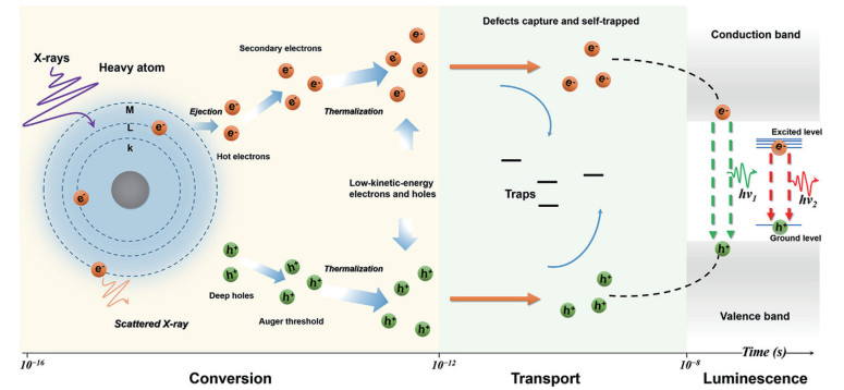
RODNYI P A, DORENBOS P, EIJK C V. Energy loss in inorganic scintillators[J]. Phys. Status Solidi B, 1995, 18715-29
WEBER M J. Scintillation: Mechanisms and new crystals[J]. Nucl. Instrum. Methods Phys. Res. Sect. A‒Accel. Spectrom. Dect. Assoc. Equip., 2004, 527(1): 9-14
ZHOU Y, CHEN J, BAKR O M, MOHAMMED O F. Metal halide perovskites for X-ray imaging scintillators and detectors[J]. ACS Energy Lett., 2021, 6(2): 739-768
doi: 10.1021/acsenergylett.0c02430
MADDALENA F, TJAHJANA L, XIE A, ARRAMEL, ZENG S W, WANG H, COQUET P, DROZDOWSKI W, DUJARDIN C, DANG C. Inorganic, organic, and perovskite halides with nanotechnology for high-light yield X- and γ-ray scintillators[J]. Crystals, 2019, 9(2): 88
doi: 10.3390/cryst9020088
NIKL M. Scintillation detectors for X-rays[J]. Meas. Sci. Technol., 2006, 17(4): R37
doi: 10.1088/0957-0233/17/4/R01
CHEN H Y, MOORE T, QI B, COLVIN D C, JELEN E K, HITCHCOCK D A, HE J, MEFFORD O T, GORE J C, ALEXIS F, ANKER J N. Monitoring pH-triggered drug release from radioluminescent nanocapsules with X-ray excited optical luminescence[J]. ACS Nano, 2013, 7(2): 1178-1187
doi: 10.1021/nn304369m
MOORE T L, WANG F L, CHEN H Y, GRIMES S W, ANKER J N, ALEXIS F. Polymer-coated radioluminescent nanoparticles for quantitative imaging of drug delivery[J]. Adv. Funct. Mater., 2014, 24(37): 5815-5823
doi: 10.1002/adfm.201400949
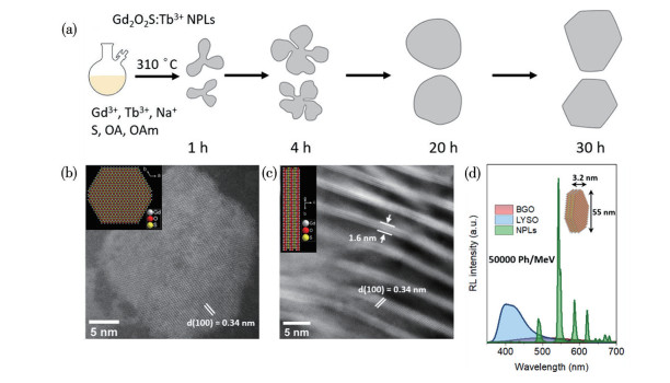
YOROV K E, NEMATULLOEV S, SAIDZHONOV B M, SKOROTETCKY M S, KARLUK A A, HASANOV B E, MIR W J, SHEIKH T, GUTIÉRREZ ARZALUZ L, PHIELEPEIT M E M, ASHRAF N, BLICK R H, MOHAMMED O F, BAYINDIR M, BAKR O M. Controlled synthesis of terbium-doped colloidal Gd2O2S nanoplatelets enables high-performance X-ray scintillators[J]. ACS Nano, 2024, 18(31): 20111-20122
doi: 10.1021/acsnano.4c01652
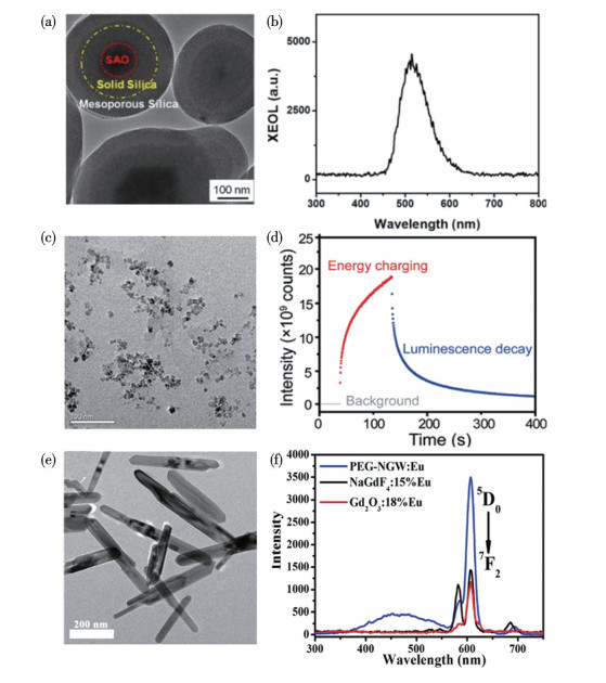
SUN C, PRATX G, CARPENTER C M, LIU H G, CHENG Z, GAMBHIR S S, XING L. Synthesis and radioluminescence of pegylated Eu3+-doped nanophosphors as bioimaging probes[J]. Adv. Mater., 2011, 23(24): 201100919
SUDHEENDRA L, DAS G K, LI C Q, STARK D, CENA J, CHERRY S, KENNEDY I M. NaGdF4∶Eu3+ nanoparticles for enhanced X-ray excited optical imaging[J]. Chem. Mater., 2014, 26(5): 1881-1888
doi: 10.1021/cm404044n
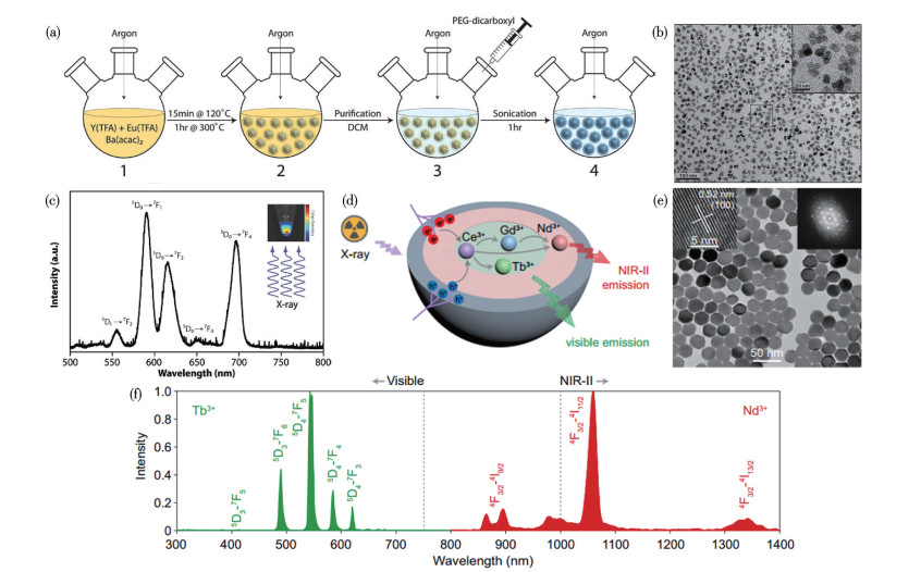
NACZYNSKI D J, SUN C, TÜRKCAN S, JENKINS C, KOH A L, IKEDA D, PRATX G, XING L. X-ray-induced shortwave infrared biomedical imaging using rare-earth nanoprobes[J]. Nano Lett., 2015, 15(1): 96-102
doi: 10.1021/nl504123r
LIANG H Y, HONG Z Z, LI S H, SONG X R, ZHANG D, CHEN Q S, LI J, YANG H H. An activatable X-ray scintillating luminescent nanoprobe for early diagnosis and progression monitoring of thrombosis in live rat[J]. Adv. Funct. Mater., 2020, 31(5): 2006353
HE L R, WANG L Y, YU X J, TANG Y Z, JIANG Z, YANG G L, LIU Z, LI W W. Full-course NIR-Ⅱ imaging-navigated fractionated photodynamic therapy of bladder tumours with X-ray-activated nanotransducers[J]. Nat. Commun., 2024, 15: 8240
doi: 10.1038/s41467-024-52607-9
CUAU L, AKL P, GAUTHERON A, HOUMEAU A, CHAPUT F, YAROMINA A, DUBOIS L, LAMBIN P, KARPATI S, PAROLA S, REZAEIFAR B, LANGLOIS J B, SI-MOHAMED S A, MONTCEL B, DOUEK P, LEROUGE F. Surface modification effect on contrast agent efficiency for X-ray based spectral photon-counting scanner/luminescence imaging: From fundamental study to in vivo proof of concept[J]. Nanoscale, 2024, 16(6): 2931-2944
doi: 10.1039/D3NR03710J
LI Y, GECEVICIUS M, QIU J. Long persistent phosphors-from fundamentals to applications[J]. Chem. Soc. Rev., 2016, 45(8): 2090-2136
doi: 10.1039/C5CS00582E
ZHANG L W, SHEN R C, TAN J, YUAN Q. Influence of doped ions on persistent luminescence materials: A review[J]. Chin. J. Struct. Chem, 2022, 41(2): 2202148-2202158
ZHENG B, FAN J, CHEN B, QIN X, WANG J, WANG F, DENG R, LIU X. Rare-earth doping in nanostructured inorganic materials[J]. Chem. Rev., 2022, 122(6): 5519-5603
doi: 10.1021/acs.chemrev.1c00644
COOPER D R, CAPOBIANCO J A, SEUNTJENS J. Radioluminescence studies of colloidal oleate-capped β-Na(Gd, Lu)F4∶Ln3+ nanoparticles (Ln=Ce, Eu, Tb)[J]. Nanoscale, 2018, 10(16): 7821-7832
doi: 10.1039/C8NR01262H
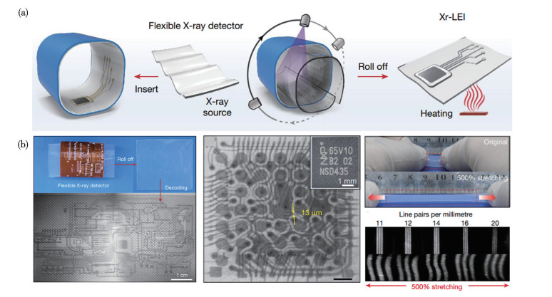
OU X Y, QIN X, HUANG B L, ZAN J, WU Q X, HONG Z Z, XIE L L, BIAN H Y, YI Z G, CHEN X F, WU Y M, SONG X R, LI J, CHEN Q S, YANG H H, LIU X G. High-resolution X-ray luminescence extension imaging[J]. Nature, 2021, 590(7846): 410-415
doi: 10.1038/s41586-021-03251-6
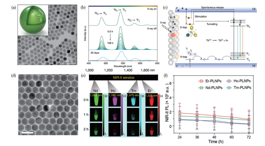
PEI P, CHEN Y, SUN C X, FAN Y, YANG Y M, LIU X, LU L F, ZHAO M Y, ZHANG H X, ZHAO D Y, LIU X G, ZHANG F. X-ray-activated persistent luminescence nanomaterials for NIR-Ⅱ imaging[J]. Nat. Nanotechnol., 2021, 16(9): 1011-1018
doi: 10.1038/s41565-021-00922-3
ZHUANG Y X, CHEN D R, CHEN W J, ZHANG W X, SU X, DENG R R, AN Z F, CHEN H M, XIE R J. X-ray-charged bright persistent luminescence in NaYF4∶Ln3+@NaYF4 nanoparticles for multidimensional optical information storage[J]. Light‒Sci. Appl., 2021, 10: 132
LEI L, WANG Y B, XU W X, YE R G, HUA Y J, DENG D G, CHEN L, PRASAD P N, XU S Q. Manipulation of time-dependent multicolour evolution of X-ray excited afterglow in lanthanide-doped fluoride nanoparticles[J]. Nat. Commun., 2022, 13: 5739
doi: 10.1038/s41467-022-33489-1
XIE F, CHEN D X, ZHANG Y, LV X L, CHEN X, SUN K N, LIANG Y J. Colloidal KLu3F10∶Tb3+ persistent luminescence nanocrystal based flexible detectors for 3D X-ray imaging[J]. J. Mater. Chem. C, 2023, 11(47): 16772-16781
doi: 10.1039/D3TC03409G
ZOU H R, ZHU W J, ZHAO J T, ZHOU S, XU S Q, LEI L. Sub-10 nm lanthanide-doped Lu6O5F8 nanoscintillators for real-time high-resolution dynamic 3D X-ray imaging[J]. Adv. Funct. Mater., 2024, 34(49): 2409156
doi: 10.1002/adfm.202409156
CHEN H M, WANG G D, CHUANG Y J, ZHEN Z P, CHEN X Y, BIDDINGER P, HAO Z L, LIU F, SHEN B Z, PAN Z W, XIE J. Nanoscintillator-mediated X-ray inducible photodynamic therapy for in vivo cancer treatment[J]. Nano Lett., 2015, 15(4): 2249-2256
doi: 10.1021/nl504044p
SONG L, LIN X H, SONG X R, CHEN S, CHEN X F, LI J, YANG H H. Repeatable deep-tissue activation of persistent luminescent nanoparticles by soft X-ray for high sensitivity long-term in vivo bioimaging[J]. Nanoscale, 2017, 9(8): 2718-2722
doi: 10.1039/C6NR09553D
GUO T, LIN Y, ZHANG W J, HONG J S, LIN R H, WU X P, LI J, LU C H, YANG H H. High-efficiency X-ray luminescence in Eu3+-activated tungstate nanoprobes for optical imaging through energy transfer sensitization[J]. Nanoscale, 2018, 10(4): 1607-1612
doi: 10.1039/C7NR06405E
ESPINOZA S, JUESTEL T, HAASE M. Colloidal LaPO4∶Gd3+ nanocrystals: X-ray induced single line UV emission[J]. Nanoscale, 2018, 10(47): 22533-22540
doi: 10.1039/C8NR06867D
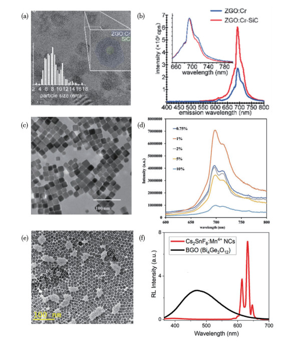
BEKE D, NARDI M V, BORTEL G, TIMPEL M, CZIGÁNY Z, PASQUALI L, CHIAPPINI A, BAIS G, RUDOLF M, ZALKA D, BIGI F, ROSSI F, BENCS L, PEKKER A, MÁRKUS B G, SALVIATI G, SADDOW S E, KAMARÁS K, SIMON F, GALI A. Enhancement of X-ray-excited red luminescence of chromium-doped zinc gallate via ultrasmall silicon carbide nanocrystals[J]. Chem. Mater., 2021, 33(7): 2457-2465
doi: 10.1021/acs.chemmater.0c04671
SHRIVASTAVA N, GUFFIE J, MOORE T L, GUZELTURK B, KUMBHAR A S, WEN J G, LUO Z P. Surface-doped zinc gallate colloidal nanoparticles exhibit pH-dependent radioluminescence with enhancement in acidic media[J]. Nano Lett., 2023, 23(14): 6482-6488
doi: 10.1021/acs.nanolett.3c01363
XUE Z L, LI X L, LI Y B, JIANG M Y, LIU H N, ZENG S J, HAO J H. X-ray-activated near-infrared persistent luminescent probe for deep-tissue and renewable in vivo bioimaging[J]. ACS Appl. Mater. Interfaces, 2017, 9(27): 22132-22142
doi: 10.1021/acsami.7b03802
CHEN Z Z, WANG L C, MANOHARAN D, LEE C L, WU L C, HUANG W T, HUANG E Y, SU C H, SHEU H S, YEH C S. Low dose of X-ray-excited long-lasting luminescent concave nanocubes in highly passive targeting deep-seated hepatic tumors[J]. Adv. Mater., 2019, 31(49): 1905087
doi: 10.1002/adma.201905087
LIU B M, ZOU R, LOU S Q, GAO Y F, MA L, WONG K L, WANG J. Low-dose X-ray-stimulated LaGaO3∶Sb, Cr near-infrared persistent luminescence nanoparticles for deep-tissue and renewable in vivo bioimaging[J]. Chem. Eng. J., 2021, 404127133
YOROV K E, MIR W J, SONG X, GUTIÉRREZ ARZALUZ L, NAPHADE R, NEMATULLOEV S, CHEN C, HUANG R W, SHAO B Y, HASANOV B E, HAN Y, MOHAMMED O F, BAKR O M. Mn4+-doped fluoride nanocrystals enable high-resolution red-emitting X-ray imaging screens[J]. ACS Mater. Lett., 2022, 4(11): 2273-2281
doi: 10.1021/acsmaterialslett.2c00746
BAWENDI M G, STEIGERWALD M L, BRUS L E. The quantum mechanics of larger semiconductor clusters ("quantum dots")[J]. Annu. Rev. Phys. Chem., 1990, 41(41): 477-496
XU G X, ZENG S W, ZHANG B T, SWIHART M T, YONG K T, PRASAD P N. New generation cadmium-free quantum dots for biophotonics and nanomedicine[J]. Chem. Rev., 2016, 116(19): 12234-12327
doi: 10.1021/acs.chemrev.6b00290
WEGNER K D, HILDEBRANDT N. Quantum dots: Bright and versatile in vitro and in vivo fluorescence imaging biosensors[J]. Chem. Soc. Rev., 2015, 44(14): 4792-4834
doi: 10.1039/C4CS00532E
MARTYNENKO I V, LITVIN A P, PURCELL MILTON F, BARANOV A V, FEDOROV A V, GUN'KO Y K. Application of semiconductor quantum dots in bioimaging and biosensing[J]. J. Mater. Chem. B, 2017, 5(33): 6701-6727
doi: 10.1039/C7TB01425B
YAO J, LI L, LI P F, YANG M. Quantum dots: From fluorescence to chemiluminescence, bioluminescence, electrochemiluminescence, and electrochemistry[J]. Nanoscale, 2017, 9(36): 13364-13383
doi: 10.1039/C7NR05233B
KANG Z T, ZHANG Y L, MENKARA H, WAGNER B K, SUMMERS C J, LAWRENCE W, NAGARKAR V. CdTe quantum dots and polymer nanocomposites for X-ray scintillation and imaging[J]. Appl. Phys. Lett., 2011, 98(18): 181914
doi: 10.1063/1.3589366
HOSSU M, LIU Z X, YAO M Z, MA L, CHEN W. X-ray luminescence of CdTe quantum dots in LaF3∶Ce/CdTe nanocomposites[J]. Appl. Phys. Lett., 2012, 100(1): 103104
GUIDELLI E J, LIGNOS I, YOO J J, LUSARDI M, BAWENDI M G, BAFFA O, JENSEN K F. Mechanistic insights and controlled synthesis of radioluminescent ZnSe quantum dots using a microfluidic reactor[J]. Chem. Mater., 2018, 30(23): 8562-8570
doi: 10.1021/acs.chemmater.8b03587
CARULLI F, COVA F, GIRONI L, MEINARDI F, VEDDA A, BROVELLI S. Stokes shift engineered Mn∶CdZnS/ZnS nanocrystals as reabsorption-free nanoscintillators in high loading polymer composites[J]. Adv. Opt. Mater., 2022, 10(13): 202200419
FANG Z H, TANG H T, YANG Z, ZHANG H, PENG Q P, YU X, ZHOU D C, QIU J B, XU X H. Transparent medium embedded with CdS quantum dots for X-ray imaging[J]. Adv. Opt. Mater., 2021, 9(24): 2101607
doi: 10.1002/adom.202101607
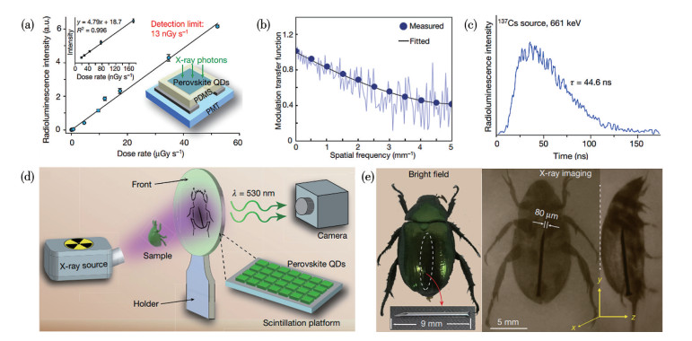
CHEN Q S, WU J, OU X Y, HUANG B L, ALMUTLAQ J, ZHUMEKENOV A A, GUAN X W, HAN S Y, LIANG L L, YI Z G, LI J, XIE X J, WANG Y, LI Y, FAN D Y, TEH D B L, ALL A H, MOHAMMED O F, BAKR O M, WU T, BETTINELLI M, YANG H H, HUANG W, LIU X G. All-inorganic perovskite nanocrystal scintillators[J]. Nature, 2018, 561(7721): 88-93
doi: 10.1038/s41586-018-0451-1
HEO J H, SHIN D H, PARK J K, KIM D H, LEE S J, IM S H. High-performance next-generation perovskite nanocrystal scintillator for nondestructive X-ray imaging[J]. Adv. Mater., 2018, 30(40): 1801743
doi: 10.1002/adma.201801743
ZHANG Y H, SUN R J, OU X Y, FU K F, CHEN Q S, DING Y C, XU L J, LIU L M, HAN Y, MALKO A V, LIU X G, YANG H H, BAKR O M, LIU H, MOHAMMED O F. Metal halide perovskite nanosheet for X-ray high-resolution scintillation imaging screens[J]. ACS Nano, 2019, 13(2): 2520-2525
doi: 10.1021/acsnano.8b09484
LI Y W, XU Y L, YAO F, LIN Q Q. Heterovalent cation-exchange of CsPbBr3 perovskite nanocrystals with enhanced stability for X-ray imaging[J]. Appl. Phys. Lett., 2023, 123(11): 111103
doi: 10.1063/5.0158665
YANG Z, YAO J S, XU L M, FAN W X, SONG J Z. Designer bright and fast CsPbBr3 perovskite nanocrystal scintillators for high-speed X-ray imaging[J]. Nat. Commun., 2024, 15: 8870
doi: 10.1038/s41467-024-53263-9
WANG Y, WANG C, MEN L, HU Q S, XIAO J W. Colloidal synthesis of hollow double perovskite nanocrystals and their applications in X-ray imaging[J]. Inorg. Chem., 2024, 63(12): 5734-5742
doi: 10.1021/acs.inorgchem.4c00280
XING G Y, CUI E D, YUAN X Y, WANG B, ZHAO Y A, TANG J F, CHEN J C, LIU J. Defects in ligand-exchange-passivated mixed- halide double perovskite nanocrystals for X-ray imaging[J]. Laser Photon. Rev., 2025, 19(5): 2401552
doi: 10.1002/lpor.202401552
GUAN L Q, SHI S, NIU X W, GUO S C, ZHAO J, JI T M, DONG H, JIA F Y, XIAO J W, SUN L D, YAN C H. All-inorganic manganese-based CsMnCl3 nanocrystals for X-ray imaging[J]. Adv. Sci., 2022, 9(18): 202201354
ZHOU J E, AN K, HE P, YANG J, ZHOU C, LUO Y, KANG W, HU W, FENG P, ZHOU M, TANG X S. Solution-processed lead-free perovskite nanocrystal scintillators for high-resolution X-ray CT imaging[J]. Adv. Opt. Mater., 2021, 9(11): 2022144
YAO Q F, ZHU M S Q, YANG Z C, SONG X R, YUAN X, ZHANG Z P, HU W P, XIE J P. Molecule-like synthesis of ligand-protected metal nanoclusters [J]. Nat. Rev. Mater., 2025, 10(2): 89-108
WANG J J, FENG L Z, SHI G Y, YANG J N, ZHANG Y D, XU H Y, SONG K H, CHEN T, ZHANG G Z, ZHENG X S, FAN F J, XIAO Z G, YAO H B. High efficiency warm-white light-emitting diodes based on copper-iodide clusters[J]. Nat. Photonics, 2024, 18(2): 200-206
doi: 10.1038/s41566-023-01340-8
ZHANG N, LI Y, HAN S Y, WEI Y, HU H, HUO R, DUAN C B, ZHANG J, HAN C M, XIE G H, XU H. Cluster light-emitting diodes containing copper iodine cube with 100% exciton utilization using host-cluster synergy[J]. Angew. Chem. ‒Int. Edit., 2023, 62(27): e202305018
doi: 10.1002/anie.202305018
OSAKADA Y, PRATX G, SUN C, SAKAMOTO M, AHMAD M, VOLOTSKOVA O, ONG Q X, TERANISHI T, HARADA Y, XING L, CUI B X. Hard X-ray-induced optical luminescence via biomolecule-directed metal clusters[J]. Chem. Commun., 2014, 50(27): 3549-3551
doi: 10.1039/C3CC48661C
LIU Z Y, JUNG K O, TAKAHATA R, SAKAMOTO M, TERANISHI T, FUJITSUKA M, PRATX G, OSAKADA Y. Hard X-ray excited optical luminescence from protein-directed Au∼20 clusters[J]. RSC Adv., 2020, 10(23): 13824-13829
doi: 10.1039/D0RA01935F
KIRAKCI K, KUBáT P, FEJFAROVá K, MARTINČíK J, NIKL M, LANG K. X-ray inducible luminescence and singlet oxygen sensitization by an octahedral molybdenum cluster compound: A new class of nanoscintillators [J]. Inorg. Chem., 2015, 55(2): 803-809
EVTUSHOK D V, MELNIKOV A R, VOROTNIKOVA N A, VOROTNIKOV Y A, RYADUN A A, KURATIEVA N V, KOZYR K V, OBEDINSKAYA N R, KRETOV E I, NOVOZHILOV I N, MIRONOV Y V, STASS D V, EFREMOVA O A, SHESTOPALOV M A. A comparative study of optical properties and X-ray induced luminescence of octahedral molybdenum and tungsten cluster complexes[J]. Dalton Trans., 2017, 46(35): 11738-11747
doi: 10.1039/C7DT01919J
KIRAKCI K, FEJFAROVÁ K, MARTINČÍK J, NIKL M, LANG K. Tetranuclear copper(Ⅰ) iodide complexes: A new class of X-ray phosphors[J]. Inorg. Chem., 2017, 56(8): 4609-4614
doi: 10.1021/acs.inorgchem.7b00240
ZHOU Y, HE T Y, YUAN P, YIN J, CHEN S L, GUTIÉRREZ ARZALUZ L, WANG L J, BAKR O M, MOHAMMED O F. Colloidal Cu4I4 clusters for high-resolution X-ray imaging scintillation screens[J]. ACS Mater. Lett., 2023, 5(8): 2002-2008
doi: 10.1021/acsmaterialslett.3c00377
LU K D, AUNG T, GUO N N, WEICHSELBAUM R, LIN W B. Nanoscale metal-organic frameworks for therapeutic, imaging, and sensing applications[J]. Adv. Mater., 2018, 30(37): 1707634
doi: 10.1002/adma.201707634
WU M X, YANG Y W. Metal-organic framework (MOF)-based drug/cargo delivery and cancer therapy[J]. Adv. Mater., 2017, 29(23): 1606134
doi: 10.1002/adma.201606134
HE C B, LIU D M, LIN W B. Nanomedicine applications of hybrid nanomaterials built from metal-ligand coordination bonds: Nanoscale metal-organic frameworks and nanoscale coordination polymers[J]. Chem. Rev., 2015, 115(19): 11079-11108
doi: 10.1021/acs.chemrev.5b00125
LAN G X, NI K Y, XU R Y, LU K D, LIN Z K, CHAN C, LIN W B. Nanoscale metal-organic layers for deeply penetrating X‑ray‑induced photodynamic therapy[J]. Angew. Chem. ‒Int. Edit., 2017, 56(40): 12102-12106
doi: 10.1002/anie.201704828
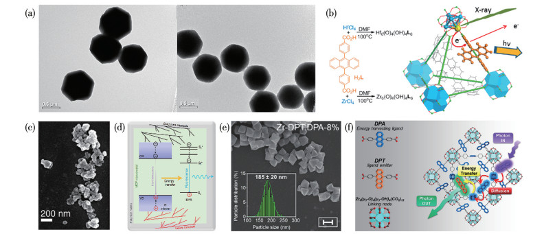
WANG C, VOLOTSKOVA O, LU K D, AHMAD M, SUN C, XING L, LIN W B. Synergistic assembly of heavy metal clusters and luminescent organic bridging ligands in metal-organic frameworks for highly efficient X-ray scintillation[J]. J. Am. Chem. Soc., 2014, 136(17): 6171-6174
doi: 10.1021/ja500671h
PEREGO J, VILLA I, PEDRINI A, PADOVANI E C, CRAPANZANO R, VEDDA A, DUJARDIN C, BEZUIDENHOUT C X, BRACCO S, SOZZANI P E, COMOTTI A, GIRONI L, BERETTA M, SALOMONI M, KRATOCHWIL N, GUNDACKER S, AUFFRAY E, MEINARDI F, MONGUZZI A. Composite fast scintillators based on high-Z fluorescent metal-organic framework nanocrystals[J]. Nat. Photonics, 2021, 15(5): 393-400
doi: 10.1038/s41566-021-00769-z
PEREGO J, BEZUIDENHOUT C X, VILLA I, COVA F, CRAPANZANO R, FRANK I, PAGANO F, KRATOCHWILL N, AUFFRAY E, BRACCO S, VEDDA A, DUJARDIN C, SOZZANI P E, MEINARDI F, COMOTTI A, MONGUZZI A. Highly luminescent scintillating hetero-ligand MOF nanocrystals with engineered Stokes shift for photonic applications[J]. Nat. Commun., 2022, 13: 3504
doi: 10.1038/s41467-022-31163-0
WANG F, DENG R R, LIU X G. Preparation of core-shell NaGdF4 nanoparticles doped with luminescent lanthanide ions to be used as upconversion-based probes[J]. Nat. Protoc., 2014, 9(7): 1634-1644
doi: 10.1038/nprot.2014.111
CHEN G Y, OHULCHANSKYY T Y, LIU S, LAW W C, WU F, SWIHART M T, ÅGREN H, PRASAD P N. Core/shell NaGdF4∶Nd3+/NaGdF4 nanocrystals with efficient near-infrared to near-infrared downconversion photoluminescence for bioimaging applications[J]. ACS Nano, 2012, 6(4): 2969-2977
doi: 10.1021/nn2042362
ZHU W J, MA W B, SU Y R, CHEN Z, CHEN X Y, MA Y G, BAI L Z, XIAO W G, LIU T Y, ZHU H M, LIU X F, LIU H F, LIU X, YANG Y. Low-dose real-time X-ray imaging with nontoxic double perovskite scintillators[J]. Light‒Sci. Appl., 2020, 9(1): 112
ZHOU J, ZHU X J, CHEN M, SUN Y, LI F Y. Water-stable NaLuF4-based upconversion nanophosphors with long-term validity for multimodal lymphatic imaging[J]. Biomaterials, 2012, 33(26): 6201-6210
doi: 10.1016/j.biomaterials.2012.05.036
JIANG M Y, XUE Z L, LI Y B, LIU H R, ZENG S J, HAO J H. A soft X-ray activated lanthanide scintillator for controllable NO release and gas-sensitized cancer therapy[J]. Nanoscale Horiz., 2020, 5(2): 268-273
doi: 10.1039/C9NH00564A
JIA T, XU J T, DONG S M, HE F, ZHONG C N, YANG G X, BI H T, XU M S, HU Y K, YANG D. Mesoporous cerium oxide-coated upconversion nanoparticles for tumor-responsive chemo-photodynamic therapy and bioimaging[J]. Chem. Sci., 2019, 10(37): 8618-8633
doi: 10.1039/C9SC01615E
DAI Y, YANG D P, YU D P, CAO C, WANG Q H, XIE S H, SHEN L, FENG W, LI F Y. Mussel-inspired polydopamine-coated lanthanide nanoparticles for NIR-Ⅱ/CT dual imaging and photothermal therapy[J]. ACS Appl. Mater. Interfaces, 2017, 9(32): 26674-26683
doi: 10.1021/acsami.7b06109
HOU B, YI L Y, HU D H, LUO Z C, GAO D Y, LI C, XING B W, WANG J W, LEE C N, ZHANG R, SHENG Z H, ZHOU B, LIU X G. A swallowable X-ray dosimeter for the real-time monitoring of radiotherapy[J]. Nat. Biomed. Eng., 2023, 7(10): 1242-1251
doi: 10.1038/s41551-023-01024-2
WIBOWO A, SHEIKH M A K, DIGUNA L J, ANANDA M B, MARSUDI M A, ARRAMEL A, ZENG S W, WONG L J, BIROWOSUTO M D. Development and challenges in perovskite scintillators for high-resolution imaging and timing applications[J]. Commun. Mater., 2023, 4(1): 21
doi: 10.1038/s43246-023-00348-5
YI L Y, HOU B, ZHAO H, LIU X G. X-ray-to-visible light-field detection through pixelated colour conversion[J]. Nature, 2023, 618(7964): 281-286
doi: 10.1038/s41586-023-05978-w
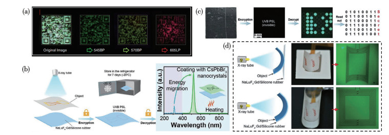
YANG Z J, ZHANG P, CHEN X F, HONG Z Z, GONG J W, OU X Y, WU Q X, LI W H, WANG X Z, XIE L L, ZHANG Z Z, YU Z Y, QIN X, TANG J, ZHANG H J, CHEN Q S, HAN S Y, YANG H H. High-confidentiality X-ray imaging encryption using prolonged imperceptible radioluminescence memory scintillators[J]. Adv. Mater., 2023, 35(52): 2309413
doi: 10.1002/adma.202309413
ZHAO X T, LI Y B, DU L M, DENG Z M, JIANG M Y, ZENG S J. Soft X-ray stimulated lanthanide@MOF nanoprobe for amplifying deep tissue synergistic photodynamic and antitumor immunotherapy[J]. Adv. Healthcare Mater., 2021, 10(21): 2101174
doi: 10.1002/adhm.202101174
MA X Q, LIN N, YANG Q, LIU P F, DING H Z, XU M J, REN F F, SHEN Z Y, HU K, MENG S S, CHEN H M. Biodegradable copper-iodide clusters modulate mitochondrial function and suppress tumor growth under ultralow‑dose X‑ray irradiation[J]. Nat. Commun., 2024, 15(1): 8092
doi: 10.1038/s41467-024-52278-6
YANG K D, YANG Y T, SUN D Q, LI S H, SONG X R, YANG H H. Designing highly UV-emitting lanthanide nanoscintillators for in vivo X-ray-activated tumor therapy[J]. Sci. China Mater., 2023, 66(10): 4090-4099
doi: 10.1007/s40843-023-2548-8
DU Z, WANG X, ZHANG X, GU Z J, FU X Y, GAN S J, FU T, XIE S T, TAN W H. X-ray-triggered carbon monoxide and manganese dioxide generation based on scintillating nanoparticles for cascade cancer radiosensitization[J]. Angew. Chem. ‒Int. Edit., 2023, 62(23): e202302525
doi: 10.1002/anie.202302525
LIU S K, LI W T, CHEN H X, ZHOU J L, DONG S M, ZANG P Y, TIAN B S, DING H, GAI S L, YANG P P, ZHAO Y L. On-demand generation of peroxynitrite from an integrated two-dimensional system for enhanced tumor therapy[J]. ACS Nano, 2022, 16(6): 8939-8953
doi: 10.1021/acsnano.1c11422
CHEN Z W, TSYTSAREV V, FINFROCK Y Z, ANTIPOVA O A, CAI Z H, ARAKAWA H, LISCHKA F W, HOOKS B M, WILTON R, WANG D Y, LIU Y, GAITAN B, TAO Y, CHEN Y, ERZURUMLU R S, YANG H H, ROZHKOVA E A. Wireless optogenetic modulation of cortical neurons enabled by radioluminescent nanoparticles[J]. ACS Nano, 2021, 15(3): 5201-5208
doi: 10.1021/acsnano.0c10436
Jian Li , Yu Zhang , Rongrong Yan , Kaiyuan Sun , Xiaoqing Liu , Zishang Liang , Yinan Jiao , Hui Bu , Xin Chen , Jinjin Zhao , Jianlin Shi . Highly Efficient, Targeted, and Traceable Perovskite Nanocrystals for Photoelectrocatalytic Oncotherapy. Acta Physico-Chimica Sinica, 2025, 41(5): 100042-0. doi: 10.1016/j.actphy.2024.100042
Zunyuan Xie , Lijin Yang , Zixiao Wan , Xiaoyu Liu , Yushan He . Exploration of the Preparation and Characterization of Nano Barium Titanate and Its Application in Inorganic Chemistry Laboratory Teaching. University Chemistry, 2024, 39(4): 62-69. doi: 10.3866/PKU.DXHX202310137
Juan Yuan , Bin Zhang , Jinping Wu , Mengfan Wang . Design of a Comprehensive Experiment on Preparation and Characterization of Cu2(Salen)2 Nanomaterials with Two Distinct Morphologies. University Chemistry, 2024, 39(10): 420-425. doi: 10.3866/PKU.DXHX202402014
Wei Li , Han Xu , Chuancan Gu , Ziyan Liu , Yan'an Li , Yan Geng . Digital Experiment on Nano-COF Materials Modulating Intracellular Ca²⁺ Concentration to Enhance Photodynamic Therapy. University Chemistry, 2026, 41(1): 354-362. doi: 10.12461/PKU.DXHX202506001
Lina Feng , Guoyu Jiang , Xiaoxia Jian , Jianguo Wang . Application of Organic Radical Materials in Biomedicine. University Chemistry, 2025, 40(4): 253-260. doi: 10.12461/PKU.DXHX202405171
Simin Fang , Wei Huang , Guanghua Yu , Cong Wei , Mingli Gao , Guangshui Li , Hongjun Tian , Wan Li . Integrating Science and Education in a Comprehensive Chemistry Design Experiment: The Preparation of Copper(I) Oxide Nanoparticles and Its Application in Dye Water Remediation. University Chemistry, 2024, 39(8): 282-289. doi: 10.3866/PKU.DXHX202401023
Bing WEI , Jianfan ZHANG , Zhe CHEN . Research progress in fine tuning of bimetallic nanocatalysts for electrocatalytic carbon dioxide reduction. Chinese Journal of Inorganic Chemistry, 2025, 41(3): 425-439. doi: 10.11862/CJIC.20240201
Renyi Shao , Khurram Abbas , Vladimir Yu. Osipov , Haimei Zhu , Yuan Li , Usama , Hong Bi . Red-emitting carbon dots prepared from Epipremnum Aureum leaves extract for biological imaging. Acta Physico-Chimica Sinica, 2026, 42(2): 100134-0. doi: 10.1016/j.actphy.2025.100134
Qiang HU , Zhiqi CHEN , Zhong CHEN , Xu WANG , Weina WU . Pyridinium-chalcone-based ClO- fluorescent probe: Preparation and biological imaging applications. Chinese Journal of Inorganic Chemistry, 2025, 41(9): 1789-1795. doi: 10.11862/CJIC.20250086
Jinlong YAN , Weina WU , Yuan WANG . A simple Schiff base probe for the fluorescent turn-on detection of hypochlorite and its biological imaging application. Chinese Journal of Inorganic Chemistry, 2024, 40(9): 1653-1660. doi: 10.11862/CJIC.20240154
Liang TANG , Jingfei NI , Kang XIAO , Xiangmei LIU . Synthesis and X-ray imaging application of lanthanide-organic complex-based scintillators. Chinese Journal of Inorganic Chemistry, 2024, 40(10): 1892-1902. doi: 10.11862/CJIC.20240139
Wenjun Zheng . Application in Inorganic Synthesis of Ionic Liquids. University Chemistry, 2024, 39(8): 163-168. doi: 10.3866/PKU.DXHX202401020
Shasha SUN , Weichun HUANG , Mengke WANG . Research progress of interface regulation strategies and applications of two‑dimensional MXenes. Chinese Journal of Inorganic Chemistry, 2025, 41(8): 1465-1482. doi: 10.11862/CJIC.20240430
Qiang MA , Yiming ZHU , Meiqi HUA , Guangyu LU , Xingdong WANG , Hailong YU , Huan PANG , Yuping LI . Research progress on metal-organic frameworks as comprehensive carrier platforms for delivering anti-glioma drugs. Chinese Journal of Inorganic Chemistry, 2026, 42(4): 657-667. doi: 10.11862/CJIC.20250250
Wei Li , Guoqiang Feng , Ze Chang . Teaching Reform of X-ray Diffraction Using Synchrotron Radiation in Materials Chemistry. University Chemistry, 2024, 39(3): 29-35. doi: 10.3866/PKU.DXHX202308060
Yuhang Zhang , Weiwei Zhao , Hongwei Liu , Junpeng Lü . Progress on Self-Powered Photodetectors Based on Low-Dimensional Materials. Acta Physico-Chimica Sinica, 2025, 41(3): 100020-0. doi: 10.3866/PKU.WHXB202310004
Yang Meiqing , Lu Wang , Haozi Lu , Yaocheng Yang , Song Liu . Recent Advances of Functional Nanomaterials for Screen-Printed Photoelectrochemical Biosensors. Acta Physico-Chimica Sinica, 2025, 41(2): 100018-0. doi: 10.3866/PKU.WHXB202310046
Zhongning Tian , Jinyuan Liu , Meng Zhang , Qianqian Jia , Mingbo Liu , Zhenjiang Li , Ting Wang , Wenjie Zhao , Dongwei Ma , Xueli Qi . Constructing selenium-vacancy-rich SiC@CoSe2−x nanocomposites to boost dipole and interfacial polarization for electromagnetic wave absorption. Acta Physico-Chimica Sinica, 2026, 42(8): 100323-0. doi: 10.1016/j.actphy.2026.100323
Hongwei Ma , Fang Zhang , Hui Ai , Niu Zhang , Shaochun Peng , Hui Li . Integrated Crystallographic Teaching with X-ray,TEM and STM. University Chemistry, 2024, 39(3): 5-17. doi: 10.3866/PKU.DXHX202308107
Hongwei Ma , Hui Li . Three Methods for Structure Determination from Powder Diffraction Data. University Chemistry, 2024, 39(3): 94-102. doi: 10.3866/PKU.DXHX202310035
 DownLoad: