Login In
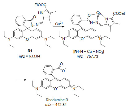
A Highly Sensitive and Selective "Off-On" Fluorescent Probe for Cu2+ Based on Rhodamine B Hydrazone
- Corresponding author: Xu Zhihong, xuzhihong1980@yahoo.com Yang Fengling, yangfengling2005@yahoo.com.cn
Figures(6)
Citation:
Lei Mengmeng, Zhou Qihang, Yang Li, Xu Zhihong, Yang Fengling. A Highly Sensitive and Selective "Off-On" Fluorescent Probe for Cu2+ Based on Rhodamine B Hydrazone[J]. Chinese Journal of Organic Chemistry,
;2020, 40(9): 2798-2803.
doi:
10.6023/cjoc202005084

Figures(6)


Wei, J. H.; Yi, J. W.; Han, M. L.; Li, B.; Liu, S.; Wu, Y. P.; Ma, L. F.; Li, D. S. A. Chem. Asian J. 2019, 14, 3694.
doi: 10.1002/asia.201900706
Wang, H.; Qin, J.; Huang, C.; Han, Y.; Xu, W.; Hou, H. Dalton Trans. 2016, 45, 12710.
doi: 10.1039/C6DT02321E
Wu, X.; Guo, Z.; Wu, Y.; Zhu, S.; James, T. D.; Zhu, W. ACS Appl. Mater. Interfaces 2013, 5, 12215.
doi: 10.1021/am404491f
Guo, Z.; Kim, G. H.; Yoon, J.; Shin, I. Nat. Protoc. 2014, 9, 1245.
doi: 10.1038/nprot.2014.086
Ma, D. L.; Wong, S. Y.; Kang, T. S.; Ng, H. P.; Han, Q. B.; Leung, C. H. Methods 2019, 168, 3.
doi: 10.1016/j.ymeth.2019.02.013
Jung, J. H.; Lee, J. H.; Shinkai, S. Chem. Soc. Rev. 2011, 40, 4464.
doi: 10.1039/c1cs15051k
Patil, P.; Ajey, K. V.; Bhat, M. P.; Sriram, G.; Yu, J.; Jung, H. Y.; Altalhi, T.; Kigga, M.; Kurkuri, M. D. ChemistrySelect 2018, 3, 11593.
doi: 10.1002/slct.201802411
Suo, F.; Chen, X.; Fang, H. Gong, Q.; Yu, C.; Yang, N. D.; Li, S.; Wu, Q.; Li, L.; Huang, W. Dyes Pigm. 2019, 170, 107639.
doi: 10.1016/j.dyepig.2019.107639
Ji, Y.; Dai, F.; Zhou, B. Free Radical Biol. Med. 2018, 129, 215.
doi: 10.1016/j.freeradbiomed.2018.09.017
Singh, N.; Paknikar, K. M.; Rajwade, J. Environ Res. 2019, 175, 367.
doi: 10.1016/j.envres.2019.05.034
Ogunkunle, C. O.; Jimoh, M. A.; Asogwa, N. T.; Viswanathan, K.; Vishwakarma, V.; Fatoba, P. O. Ecotoxicol. Environ. Saf. 2018, 155, 86.
doi: 10.1016/j.ecoenv.2018.02.070
Donnelly, P. S.; Xiao, Z.; Wedd, A. G. Curr. Opin. Chem. Biol. 2007, 11, 128.
doi: 10.1016/j.cbpa.2007.01.678
Squitti, R.; Ghidoni, R.; Simonelli, I.; Ivanova, I. D.; Colabufo, N. A.; Zuin, M.; Benussi, L.; Binetti, G.; Cassetta, E.; Rongioletti, M.; Siotto, M. J. Trace Elem. Med. Biol. 2018, 45, 181.
doi: 10.1016/j.jtemb.2017.11.005
Gardner, B.; Dieriks, B. V.; Cameron, S.; Mendis, L. H. S.; Turner, C.; Faull, R. L. M.; Curtis, M. A. Sci. Rep. 2017, 7, 10454.
doi: 10.1038/s41598-017-10659-6
Ren, D.; Liu, Y.; Liu, X.; Li, Z.; Li, H.; Yang, X. F. Sens. Actuators. B 2018, 255, 2321.
doi: 10.1016/j.snb.2017.09.048
Yoon, J. W.; Chang, M. J.; Hong, S.; Lee, M. H. Tetrahedron Lett. 2017, 58, 3887.
doi: 10.1016/j.tetlet.2017.08.071
Jiao, Y.; Zhou, L.; He, H.; Yin, J.; Gao, Q.; Wei, J.; Duan, C.; Peng, X. Talanta 2018, 184, 143.
doi: 10.1016/j.talanta.2018.01.073
Jia, X.; Xiao, Z.; Hui, P.; Liu, C.; Wang, Q.; Qiu, X. He, S.; Zeng, X.; Zhao, L. Dyes Pigm. 2019, 160, 633.
doi: 10.1016/j.dyepig.2018.08.060
Kang, H.; Fan, C.; Xu, H.; Pu, G.; Liu, S. Tetrahedron 2018, 74, 4390.
doi: 10.1016/j.tet.2018.07.002
Zhang, J. F.; Zhou, Y.; Yoon, J.; Kim, Y.; Kim, S. J.; Kim, J. S. Org Lett. 2010, 12, 3852.
doi: 10.1021/ol101535s
Yang, Z.; She, M.; Zhang, J.; Chen, X.; Huang, Y.; Zhu, H.; Liu, P.; Li, J.; Shi Z. Sens. Actuators. B 2013, 176, 482.
doi: 10.1016/j.snb.2012.07.035
Yu, M.; Yuan, R.; Shi, C.; Zhou, W.; Wei, L.; Li, Z. Dyes Pigm. 2013, 99, 887.
doi: 10.1016/j.dyepig.2013.07.030
Goswami, S.; Sen, D.; Das, A. K.; Das, N. K.; Aich, K.; Fun, H. K.; Quah, C. K.; Maity, A. K.; Saha, P. Sensors. Actuators. B 2013, 183, 518.
doi: 10.1016/j.snb.2013.04.005
Kar, C.; Adhikari, M. D.; Ramesh, A.; Das, G. Inorg. Chem. 2013, 52, 743.
doi: 10.1021/ic301872q
Xiong, K.; Chen, J. G. Catalysis Today. 2018, 339, 289.
Xu, Q.; Guo, Z. J. East China Univ. Sci. Technol. 2019, 45, 357.
Yi, X.; Li, G.; Huang, L.; Chu, Y.; Liu, Z. Q.; Xia, H.; Zheng, A.; Deng, F. J. Phys. Chem. C. 2017, 121, 3887.
doi: 10.1021/acs.jpcc.6b11518
Jr, P. A.; Liu, Y.; Palacios, M. A.; Minami, T. Wang, Z.; Nishiyabu, R. Chem.-Eur. J. 2013, 19, 8497.
doi: 10.1002/chem.201204188
Wang, Y.; Wu, H.; Wu, W. N.; Li, S. J.; Xu, Z. H.; Xu, Z. Q. Fan, Y. C.; Zhao, X. L.; Liu, B. Z. Sens. Actuators. B 2018, 260, 106.
doi: 10.1016/j.snb.2017.12.201
Moubaraki, R.; Li, B.; Murray, K. S.; Brooker, S. Eur. J. Inorg. Chem. 2009, 19, 2851.
Wang, Y.; Chang, H. Q.; Wu, W. N.; Peng, W. B.; Yan, Y. F. He, C. M.; Chen, T. T.; Zhao, X. L.; Xu, Z. Q. Sensors. Actuators B:Chem. 2016, 228, 395.
doi: 10.1016/j.snb.2016.01.052
Li, M.; Lv, H.; Luo, J. Z.; Miao, J. Y.; Zhao, B. X. Sens. Actuators. B 2013, 188, 1235.
doi: 10.1016/j.snb.2013.08.030
Ge, F.; Ye, H.; Luo, J. Z.; Wang, S.; Sun, Y. J.; Zhao, B. X.; Miao, J. Y. Sens. Actuators. B 2013, 181, 215.
doi: 10.1016/j.snb.2013.01.048
Huang, K.; Jiao, X.; Liu, C.; Wang, Q.; Qiu, X.; He, S. Zhao, L.; Zeng, X. Dyes Pigm. 2017, 145, 561.
doi: 10.1016/j.dyepig.2017.06.047
Artesani, A.; Binet, L.; Tana, F.; Comelli, D.; Nardo, L. D.; Nevin, A.; Touati, N.; Valentini, G.; Gourier, D. Microchem. J. 2019, 151, 104256.
doi: 10.1016/j.microc.2019.104256
Pressman, J. G.; Richardson, S. D.; Speth, T. F.; Miltner, R. J. Environ. Sci. Technol. 2010, 44, 7184.
doi: 10.1021/es9039314
Xu, Z.; Wang, H.; Hou, X.; Xu, W.; Xiang, T.; Wu, C. Sens. Actuators. B 2014, 201, 469.
doi: 10.1016/j.snb.2014.05.026
Dujols, V.; Ford, F.; Czarnik, A. W. J. Am. Chem. Soc. 1997, 119, 7386.
doi: 10.1021/ja971221g
Kim, H.; Rao, B. A.; Jeon, J. W.; Mallick, S.; Kang, S. M.; Choi, J. S.; Lee, C. S.; Son, Y. A. Sens. Actuators. B 2015, 210, 173.
doi: 10.1016/j.snb.2014.12.100
Ye, X. P.; Zhu, T. F.; Wu, W. N.; Ma, T. L.; Xu, J.; Zhang, Z. P.; Wang, Y.; Jia, L. Inorg. Chem. Commun. 2014, 47, 60.
doi: 10.1016/j.inoche.2014.07.022
Yuan ZHU , Xiaoda ZHANG , Shasha WANG , Peng WEI , Tao YI . Conditionally restricted fluorescent probe for Fe3+ and Cu2+ based on the naphthalimide structure. Chinese Journal of Inorganic Chemistry, 2025, 41(1): 183-192. doi: 10.11862/CJIC.20240232
Meirong HAN , Xiaoyang WEI , Sisi FENG , Yuting BAI . A zinc-based metal-organic framework for fluorescence detection of trace Cu2+. Chinese Journal of Inorganic Chemistry, 2024, 40(8): 1603-1614. doi: 10.11862/CJIC.20240150
Wei GAO , Meiqi SONG , Xuan REN , Jianliang BAI , Jing SU , Jianlong MA , Zhijun WANG . A self-calibrating fluorescent probe for the selective detection and bioimaging of HClO. Chinese Journal of Inorganic Chemistry, 2025, 41(6): 1173-1182. doi: 10.11862/CJIC.20250112
Lei ZHANG , Cheng HE , Yang JIAO . An azo-based fluorescent probe for the detection of hypoxic tumor cells. Chinese Journal of Inorganic Chemistry, 2025, 41(6): 1162-1172. doi: 10.11862/CJIC.20250081
Jun LUO , Baoshu LIU , Yunchang ZHANG , Bingkai WANG , Beibei GUO , Lan SHE , Tianheng CHEN . Europium(Ⅲ) metal-organic framework as a fluorescent probe for selectively and sensitively sensing Pb2+ in aqueous solution. Chinese Journal of Inorganic Chemistry, 2024, 40(12): 2438-2444. doi: 10.11862/CJIC.20240240
Yue Li , Qianyu Ding , Wansheng Liu , Yimeng Sun , Liyao Liu , Ye Zou , Yutao Cui , Jia Zhu , Chongan Di , Daoben Zhu . Bipyridine-bridged Φ-shaped cyclo[8]thiophene[2]pyrrole: Synthesis and fluorescence properties. Chinese Chemical Letters, 2026, 37(2): 111989-. doi: 10.1016/j.cclet.2025.111989
Shuwen SUN , Gaofeng WANG . Design and synthesis of a Zn(Ⅱ)-based coordination polymer as a fluorescent probe for trace monitoring 2, 4, 6-trinitrophenol. Chinese Journal of Inorganic Chemistry, 2025, 41(4): 753-760. doi: 10.11862/CJIC.20240399
Meitong Wu , Ke Wu , Shumin Feng , Li Xu , Mi Lei , Jianmei Chen , Shuang Li , Mian Qin , Dahui Liu , Guoqiang Feng . A NIR and ratiometric fluorescent probe for quantitative detection of SO2 derivatives in Chinese medicinal materials and bioimaging in vivo. Chinese Chemical Letters, 2026, 37(1): 110979-. doi: 10.1016/j.cclet.2025.110979
Kairong Yang , Bingbing Zheng , Fapu Wu , Bijia Zhou , Lijun Li , Hu Xiong . H2S-activated near-infrared fluorescent probe for detecting colon cancer and rapid fecal analysis. Chinese Chemical Letters, 2026, 37(4): 111235-. doi: 10.1016/j.cclet.2025.111235
Yaheng Li , Weijiang He , Yuncong Chen , Zijian Guo . A BODIPY-based ratiometric fluorescent probe for imaging of Zn2+ in ferroptosis. Chinese Chemical Letters, 2026, 37(5): 111337-. doi: 10.1016/j.cclet.2025.111337
Xian XIA , Qin SHI , Wanyi SU , Qingjun XUE , Honghui PAN , Xixiang LIU , Chuanqi ZHAO . In-situ synthesis of Bi2O3@BiVO4 composite via solvothermal method and its adsorption performance for rhodamine B in water. Chinese Journal of Inorganic Chemistry, 2026, 42(5): 1096-1112. doi: 10.11862/CJIC.20250311
Yudi Cheng , Xiao Wang , Jiao Chen , Zihan Zhang , Jiadong Ou , Mengyao She , Fulin Chen , Jianli Li . A near-infrared fluorescent probe for visualizing transformation pathway of Cys/Hcy and H2S and its applications in living system. Chinese Chemical Letters, 2024, 35(5): 109156-. doi: 10.1016/j.cclet.2023.109156
Bangdi GE , Xiaowei SONG , Zhiqiang LIANG . A bifunctional three-dimensional Eu-MOF fluorescent probe for highly sensitive detection of 2, 4, 6-trinitrophenol and tetracycline. Chinese Journal of Inorganic Chemistry, 2025, 41(10): 2165-2174. doi: 10.11862/CJIC.20250190
Anjing Liao , Wei Sun , Yaming Liu , Han Yan , Zhi Xia , Jian Wu . Pyrrole and pyrrolidine analogs: The promising scaffold in discovery of pesticides. Chinese Chemical Letters, 2025, 36(3): 110094-. doi: 10.1016/j.cclet.2024.110094
Jinlong YAN , Weina WU , Yuan WANG . A simple Schiff base probe for the fluorescent turn-on detection of hypochlorite and its biological imaging application. Chinese Journal of Inorganic Chemistry, 2024, 40(9): 1653-1660. doi: 10.11862/CJIC.20240154
Yu SU , Xinlian FAN , Yao YIN , Lin WANG . From synthesis to application: Development and prospects of InP quantum dots. Chinese Journal of Inorganic Chemistry, 2024, 40(11): 2105-2123. doi: 10.11862/CJIC.20240126
Yingpeng ZHANG , Xingxing LI , Yunshang YANG , Zhidong TENG . A pyrazole-based turn-off fluorescent probe for visual detection of hydrazine. Chinese Journal of Inorganic Chemistry, 2025, 41(7): 1301-1308. doi: 10.11862/CJIC.20250064
Yuting DU , Jing YUAN , Peiyao DENG . Synthesis and application of a fluorescent probe for the detection of reduced glutathione. Chinese Journal of Inorganic Chemistry, 2025, 41(7): 1331-1337. doi: 10.11862/CJIC.20240461
Pengli GUAN , Renhu BAI , Xiuling SUN , Bin LIU . Trianiline-derived aggregation-induced emission luminogen probe for lipase detection and cell imaging. Chinese Journal of Inorganic Chemistry, 2025, 41(9): 1817-1826. doi: 10.11862/CJIC.20250058
Qiang HU , Zhiqi CHEN , Zhong CHEN , Xu WANG , Weina WU . Pyridinium-chalcone-based ClO- fluorescent probe: Preparation and biological imaging applications. Chinese Journal of Inorganic Chemistry, 2025, 41(9): 1789-1795. doi: 10.11862/CJIC.20250086
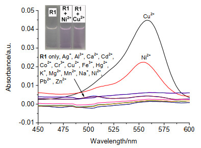
Metal ions: Ag+, Al3+, Ca2+, Cd2+, Co2+, Cr3+, Cu2+, Fe3+, Hg2+, K+, Mg2+, Mn2+, Na+, Ni2+, Pb2+, Zn2+ and blank. The inset shows the change of the color with the addition of Cu2+/Ni2+ to R1 in buffered solution
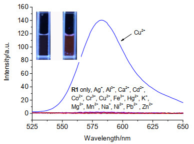
Metal ions: Ag+, Al3+, Ca2+, Cd2+, Co2+, Cr3+, Cu2+, Fe3+, Hg2+, K+, Mg2+, Mn2+, Na+, Ni2+, Pb2+ and Zn2+ and blank. The inset shows the fluorescence color of R1 with Cu2+
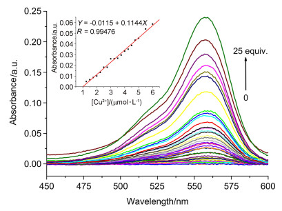
The inset shows the absorbance at 560 nm as a function of Cu2+ concentration (1.25~6.0 μmol•L-1)
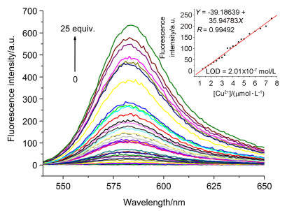
The inset shows the fluorescence intensity at 585 nm as a function of Cu2+ concentration (1.25~7.50 μmol•L-1)
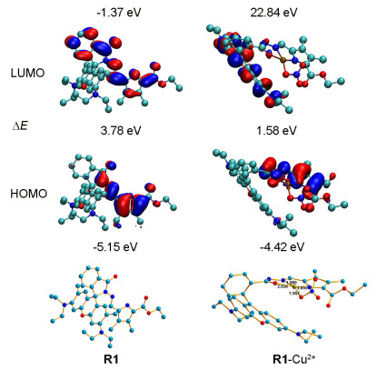
H atoms are omitted for clarity
 DownLoad: