Login In
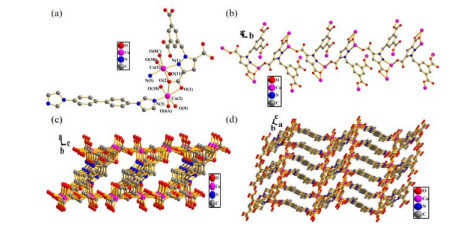
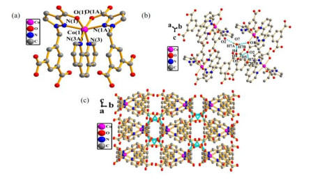
Two Co(Ⅱ) Complexes Constructed from 1-(3, 5-Dicarboxybenzyl)-3, 5-pyrazole Dicarboxylic Acid: Syntheses, Structures and Magnetic Properties
- Corresponding author: Ji-Jiang WANG, yadxwjj@126.com Long TANG, ydtanglong@163.com
Figures(7)
Citation:
Lao-Bang WANG, Ji-Jiang WANG, Long TANG, Xiao WANG, Xiang-Yang HOU, Er-Lin YUE, Yu-Qi ZHANG. Two Co(Ⅱ) Complexes Constructed from 1-(3, 5-Dicarboxybenzyl)-3, 5-pyrazole Dicarboxylic Acid: Syntheses, Structures and Magnetic Properties[J]. Chinese Journal of Structural Chemistry,
;2021, 40(7): 885-891.
doi:
10.14102/j.cnki.0254-5861.2011-3071

Figures(7)


Rouffet, M.; de Oliveira, C. A. F.; Udi, Y.; Agrawal, A.; Sagi, I.; McCammon, J. A.; Cohen, S. M. From sensors to silencers: quinoline- and benzimidazole-sulfonamides as inhibitors for zinc proteases. J. Am. Chem. Soc. 2010, 132, 8232–8233.
doi: 10.1021/ja101088j
Liu, H. Y.; Wu, H.; Ma, J. F.; Liu, Y. Y.; Liu, B.; Yang, J. Syntheses, structures, and photoluminescence of zinc(Ⅱ) coordination polymers based on carboxylates and flexible bis-[(pyridyl)-benzimidazole] ligands. Cryst. Growth Des. 2010, 10, 4795–4805.
doi: 10.1021/cg100688z
Xue, F.; Kumar, P.; Xu, W. Q.; Mkhoyan, K. A.; Tsapatsis, M. Direct synthesis of 7 nm-thick zinc(Ⅱ)-benzimidazole-acetate metal-organic framework nanosheets. Chem. Mater. 2018, 30, 69–73.
doi: 10.1021/acs.chemmater.7b04083
Yang, F.; Xu, G.; Dou, Y. B.; Wang, B.; Zhang, H.; Wu, H.; Zhou, W.; Li, J. R.; Chen, B. L. A flexible metal-organic framework with a high density of sulfonic acid sites for proton conduction. Nat. Energy 2017, 2, 877−883.
doi: 10.1038/s41560-017-0018-7
Wang, Y. L.; Han, C. B.; Zhang, Y. Q.; Liu, Q. Y.; Liu, C. M.; Yin, S. G. Fine-tuning ligand to modulate the magnetic anisotropy in a carboxylate-bridged Dy2 single-molecule magnets system. Inorg. Chem. 2016, 55, 5578–5584.
doi: 10.1021/acs.inorgchem.6b00653
Horike, S.; Umeyama, D.; Kitagawa, S. Ion conductivity and transport by porous coordination polymers and metal-organic frameworks. \ Chem. Res. 2013, 46, 2376–2384.
doi: 10.1021/ar300291s
Zhao, M.; Ou, S.; Wu, C. D. Porous metal-organic frameworks for heterogeneous biomimetic catalysis. Chem. Res. 2014, 47, 1199–1207.
doi: 10.1021/ar400265x
Kitagawa, S.; Kitaura, R.; Noro, R. Functional porous coordination polymers. Angew. Chem. Int. Ed. 2004, 43, 2334–2375.
doi: 10.1002/anie.200300610
He, Y. B.; Zhou, W.; Qian, G. D.; Chen, B. L. Methane storage in metal-organic frameworks. Chem. Soc. Rev. 2014, 43, 5657−5678.
doi: 10.1039/C4CS00032C
Lustig, W. P.; Mukherjee, S.; Rudd, N. D.; Desai, A. V.; Li, J.; Ghosh, S. K. Metal-organic frameworks: functional luminescent and photonic materials for sensing applications. Chem. Soc. Rev. 2017, 46, 3242–3285.
doi: 10.1039/C6CS00930A
Lin, Z. T.; Wang, Y. L.; Liu, Q. Y. Crystal structure and luminescence of a Cd(Ⅱ) complex based on the 3, 3΄, 5, 5΄-tetrafluorobiphenyl-4, 4΄-dicarboxylate and adenine ligands. Chin. J. Struct. Chem. 2020, 11, 2041−2045.
Zhang, N.; Guo, Y. H.; Yu, Y. Z.; Wang, Z.; Niu, Y. S.; Wu, X. L. Solvothermal synthesis, crystal Structure and luminescence property of a 1D silver(Ⅰ) coordination polymer. Chin. J. Struct. Chem. 2020, 11, 2009−2015.
Verma, P.; Singh, U. P.; Butcher, R. Luminescent metal organic frameworks for sensing and gas adsorption studies. CrystEngComm. 2019, 21, 5470–5481.
doi: 10.1039/C9CE00732F
Wang, J. J.; Cao, Z.; Wang, X.; Tang, L.; Hou, X. Y.; Ju, P.; Ren, Y. X.; Chen, X. L.; Zhang, Y. Q. A novel 3D Cd(Ⅱ) coordination polymer generated via in situ ligand synthesis involving C–O esterbond formation. RSC Adv. 2019, 9, 307–312
doi: 10.1039/C8RA06112B
Liu, C. B.; Li, Q.; Wang, X.; Che, G. B.; Zhang, X. J. A series of lanthanide(Ⅲ) coordination polymers derived via in situ hydrothermal decarboxylation of quinoline-2, 3-dicarboxylic acid. Inorg. Chem. Commun. 2014, 39, 56–60.
doi: 10.1016/j.inoche.2013.10.050
Yang, A. H.; Zou, J. Y.; Wang, W. M.; Shi, X. Y.; Gao, H. L.; Cui, J. Z.; Zhao, B. Two three-dimensional lanthanide frameworks exhibiting luminescence increases upon dehydration and novel water layer involving in situ decarboxylation. Inorg. Chem. 2014, 53, 7092–7100.
doi: 10.1021/ic402803s
Sheldrick, G. M. SHELXS-2014/7 and SHELXL-2014/7 program for solution and refinement of crystal structures. Institute for Inorganic Chemistry. University of Göttingen, Göttingen, Germany 2014.
Sheldrick, G. M. Crystal structure refinement with SHELXL. Acta Cryst. 2015, C71, 3–8.
Ishida, T.; Kawakami, T.; Mitsubori, S.; Nogami, T.; Yamaguchi, K.; Iwamura, H. Antiferromagnetic coupling of transition metal spins across pyrimidine and pyrazine bridges in dinuclear manganese(Ⅱ), cobalt(Ⅱ), nickel(Ⅱ) and copper(Ⅱ) 1, 1, 1, 5, 5, 5-hexafluoropentane-2, 4-dionate complexes. J. Chem. Soc. Dalton Trans. 2002, 3177–3186.
Ya-Xuan Xue , Han Xu , Jia-Nan Chen , Hai-Quan Tian , Tao Jia , Wei-Dong Liu , Chong-Yang Li , La-Sheng Long , Lan-Sun Zheng , Xiang-Jian Kong . Chiral Ln3Co5 clusters with geometry-dependent chiroptical and magneto-optical properties. Chinese Journal of Structural Chemistry, 2026, 45(1): 100764-100764. doi: 10.1016/j.cjsc.2025.100764
Long TANG , Yaxin BIAN , Luyuan CHEN , Xiangyang HOU , Xiao WANG , Jijiang WANG . Syntheses, structures, and properties of three coordination polymers based on 5-ethylpyridine-2,3-dicarboxylic acid and N-containing ligands. Chinese Journal of Inorganic Chemistry, 2024, 40(10): 1975-1985. doi: 10.11862/CJIC.20240180
Kun Wang , Tianxue Gong , Yaohuang Huang , Boyang Han , Hanxiao Yang , Pavlo O. Dral , Weiwei Fang . Bornylimidazo[1,5–a]pyridin-3-ylidene allylic Pd catalyst with optimal electronic and steric properties for synthesis of 3,3′-disubstituted oxindoles. Chinese Chemical Letters, 2025, 36(7): 110539-. doi: 10.1016/j.cclet.2024.110539
Jiajia Zhuang , Chunyu Cui , Changjiang Li , Gang Luo , Jiaping Tong , Di Sun . Counter-ion effect to the Ising-type magnetic anisotropy and magnetic relaxation in trigonal bipyramidal Co(Ⅱ) complexes. Chinese Chemical Letters, 2025, 36(7): 110091-. doi: 10.1016/j.cclet.2024.110091
Yanjie Li , Chaoqun Qu , Siqi Meng , Jiaqi Hu , Ze Gao , Hongji Xu , Rui Gao , Ming Feng . Revealing electronic state evolution of Co(Ⅱ)/Co(Ⅲ) in CoO (111) plane during OER process through magnetic measurement. Chinese Chemical Letters, 2025, 36(3): 109872-. doi: 10.1016/j.cclet.2024.109872
Pu ZHANG , Youzhu YU , Yuhua GUO , Zhongyuan ZHOU . Syntheses and photocatalytic CO2 reduction properties of heterometallic Ni/Sn and Co/Sn oxo clusters. Chinese Journal of Inorganic Chemistry, 2026, 42(5): 1039-1047. doi: 10.11862/CJIC.20250353
Zhao-Bo Hu , Ling-Ao Gui , Long-He Li , Tong-Tong Xiao , Adam T. Hand , Pagnareach Tin , Mykhaylo Ozerov , Yan Peng , Zhongwen Ouyang , Zhenxing Wang , Zi-Ling Xue , You Song . CoⅡ single-ion magnet and its multi-dimensional aggregations: Influence of the structural rigidity on magnetic relaxation process. Chinese Chemical Letters, 2025, 36(2): 109600-. doi: 10.1016/j.cclet.2024.109600
Xiaxia LIU , Xiaofang MA , Luxia GUO , Xianda HAN , Sisi FENG . Structure and magnetic properties of Mn(Ⅱ) coordination polymers regulated by N-auxiliary ligands. Chinese Journal of Inorganic Chemistry, 2025, 41(3): 587-596. doi: 10.11862/CJIC.20240269
Chenxi Shang , Boxuan Lu , Chongbei Wu , Shuqing Zhou , Luyan Shi , Tayirjan Taylor Isimjan , Xiulin Yang . Inducing electronic rearrangement through Co3B-Mo2B5 catalysts: Efficient dual-function catalysis for NaBH4 hydrolysis and 4-nitrophenol reduction. Chinese Chemical Letters, 2025, 36(9): 111152-. doi: 10.1016/j.cclet.2025.111152
Weizhong LING , Jingyi LIN , Jianglin ZHU , Yuyi LIANG , Shanshan DAI , Yu LI . Syntheses, structures, and catalytic performances of complexes with 4,4′-dihydroxy-[1,1′-biphenyl]-3,3′-dicarboxylic acid ligands. Chinese Journal of Inorganic Chemistry, 2026, 42(1): 152-160. doi: 10.11862/CJIC.20250204
Shuwen SUN , Gaofeng WANG . Two cadmium coordination polymers constructed by varying Ⅴ-shaped co-ligands: Syntheses, structures, and fluorescence properties. Chinese Journal of Inorganic Chemistry, 2024, 40(3): 613-620. doi: 10.11862/CJIC.20230368
Zhengzheng LIU , Pengyun ZHANG , Chengri WANG , Shengli HUANG , Guoyu YANG . Synthesis, structure, and electrochemical properties of a sandwich-type {Co6}-cluster-added germanotungstate. Chinese Journal of Inorganic Chemistry, 2024, 40(6): 1173-1179. doi: 10.11862/CJIC.20240039
Ke Xu , Shulai Lei , Panshuo Wang , Weiyi Wang , Yuan Feng , Junsheng Feng . Unraveling the microscopic origin of out of plane magnetic anisotropy in Ⅵ3. Chinese Chemical Letters, 2025, 36(8): 110257-. doi: 10.1016/j.cclet.2024.110257
Zhenghua ZHAO , Qin ZHANG , Yufeng LIU , Zifa SHI , Jinzhong GU . Syntheses, crystal structures, catalytic and anti-wear properties of nickel(Ⅱ) and zinc(Ⅱ) coordination polymers based on 5-(2-carboxyphenyl)nicotinic acid. Chinese Journal of Inorganic Chemistry, 2024, 40(3): 621-628. doi: 10.11862/CJIC.20230342
Kaimin WANG , Xiong GU , Na DENG , Hongmei YU , Yanqin YE , Yulu MA . Synthesis, structure, fluorescence properties, and Hirshfeld surface analysis of three Zn(Ⅱ)/Cu(Ⅱ) complexes based on 5-(dimethylamino) isophthalic acid. Chinese Journal of Inorganic Chemistry, 2024, 40(7): 1397-1408. doi: 10.11862/CJIC.20240009
Weizhong LING , Xiangyun CHEN , Wenjing LIU , Yingkai HUANG , Yu LI . Syntheses, crystal structures, and catalytic properties of three zinc(Ⅱ), cobalt(Ⅱ) and nickel(Ⅱ) coordination polymers constructed from 5-(4-carboxyphenoxy)nicotinic acid. Chinese Journal of Inorganic Chemistry, 2024, 40(9): 1803-1810. doi: 10.11862/CJIC.20240068
Jing FENG , Renshu WANG , Hu WANG , Hailong LIU . Co(Ⅱ) and Ni(Ⅱ) complexes of 3,3-diphenylpropionic acid and 2,2′-dipyridylamine: Structures and biological activities. Chinese Journal of Inorganic Chemistry, 2026, 42(3): 617-631. doi: 10.11862/CJIC.20250265
Feng-Fan Yang , Yin-Kang Ding , Lin-Kai Wu , Jiayue Tian , Shuai Dou , Wenjing Wang , Linfeng Liang . A 1,3,5-triazine μ3-bridged neutral Cu(Ⅰ) framework with enhanced stability and CO2 capture selectivity. Chinese Chemical Letters, 2025, 36(12): 110550-. doi: 10.1016/j.cclet.2024.110550
Xiaofan ZHANG , Yu DUAN , Meijie SHI , Nan LU , Renhong LI , Xiaoqing YAN . Z-scheme Co3O4/BiOBr heterojunction for efficient photoreduction CO2 reduction. Chinese Journal of Inorganic Chemistry, 2025, 41(9): 1878-1888. doi: 10.11862/CJIC.20250079
Weichen WANG , Chunhua GONG , Junyong ZHANG , Yanfeng BI , Hao XU , Jingli XIE . Construction of two metal-organic frameworks by rigid bis(triazole) and carboxylate mixed-ligands and their catalytic properties for CO2 cycloaddition reaction. Chinese Journal of Inorganic Chemistry, 2024, 40(7): 1377-1386. doi: 10.11862/CJIC.20230415
 DownLoad: