Citation: Yang YANG, Jian-Ping YONG, Zacchaeus Olatunde OLAGOKE, Can-Zhong LU. First Isolation and Confirmation of Sterol Based on β-sitosterol Skeleton from the Leaves of Podocarpusnagi Planted in Fujian, Preliminary in vitro Anticancer Activity and the Crystal Structure[J]. Chinese Journal of Structural Chemistry, ;2021, 40(5): 653-658. doi: 10.14102/j.cnki.0254–5861.2011–2981 shu

First Isolation and Confirmation of Sterol Based on β-sitosterol Skeleton from the Leaves of Podocarpusnagi Planted in Fujian, Preliminary in vitro Anticancer Activity and the Crystal Structure

  • Corresponding author: Jian-Ping YONG, jpyong@fjirsm.ac.cn Can-Zhong LU, czlu@fjirsm.ac.cn
  • Received Date: 14 September 2020
    Accepted Date: 23 November 2020

    Fund Project: the National Natural Science Foundation of China 21875252Self-created Area Project of Major Science Technology Innovation Platform of Xiamen 3502ZCQ20171002

Figures(3)

  • The crystal of (24R)-3β, 5α-dihydroxy-24-ethyl-5α-cholestan-6-one (1) was isolated from the leaves of P. nagi planted in Fujian for the first time with the same skeleton as 26, 27-dinorcholest-5-en-3-β-ol (2) and β-sitosterol (3) that have been reported before. Compound 1 crystallizes in monoclinic, space group P21 with a = 10.8482(4), b = 7.3671(3), c = 33.7860(15) Å, β = 93.103(4)°, V = 2696.21(19) Å3, Z = 75, Mr = 446.70, ρcalc = 1.145 g/cm3, F(000) = 1032, μ = 0.572 mm–1, GOOF = 1.034, the final R = 0.0467 and wR = 0.1129 for 6989 observed reflections with I > 2σ(I). Compound 2 was selected to evaluate for their preliminary in vitro anticancer activity against four cancer cell lines for the first time. The results showed that compound 2 exhibited great inhibition against gastric cancer, breast cancer MCF-7, lung cancer A549 and Helacell lines with the inhibitions of 89.16% ±1.17, 97.02%±0.53, 47.20% ±2.58 and 36.89%±1.22 respectively at the concentration of 1.4×10⁻² M, which means that we found the new anticancer compound in this plant medicine.
  • Podocarpusnagi (P. nagi, named Zhubai in Chinese) is widely distributed in south districts of Yangtze River, such as Fujian, Hunan, Guangxi, Guangdong, etc. This plant contains different kinds of biological compounds (such as volatile oil, flavonoids, steroids, sugar and glycosides, lactones and so on) and exhibits a wide spectrum of biological activities like hemostasis, bone setting, anti-bacterial, anti-tumor, antiviral, antioxidant and detume-scence activities[1]. According to the folk records of the Yao Nationality, P. nagi has ever been used to treat trauma, stop-bleeding, fractures, knife wounds, gunshot wounds, body odor, eye diseases, colds, and so forth. The fresh bark or root of P. nagi was also used to treat the rheumatoid arthritis[2-4]. Some work about the chemical components and biological activities of P.nagi has been reported: Ye Yang and XuYaming's groups isolated Podocarpus nagilactones from P. nagiplanted in Guangdong province and evaluated their biological activity. The results showed that most of them exhibited higher antitumor activity[5, 6]. Chen Yegao's group isolated several bioflavo- noids and few steroids from the leaves of P. nagi grown in Yunnan[7]. However, P. nagi was also distributed in Nan- ping of Fujian province. In recent years, a large scale of P. nagi was planted in Yangli town of Fujian province. Our research group has extracted the essential oil from its fruits, confirmed its chemical components, and evaluated its biological activities. The results showed this oil contains many active components: such as abundant unsaturated fatty acids, flavonoids, β-vanillin, vitamin E and essential microelements for human body; the biological evaluation results showed that it exhibited higher anti-oxidant[8]. We checked the published papers and some old records and found that there are some differences about the P. nagi grown in Guangdong, Yunnan and Fujian. To the best of our knowledge, there are no reports about the chemical components of P. nagi planted in Fujian, so our research group took the lead to isolate the chemical components from the leaves of P. nagi planted in Fujian.

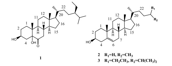
    In this work, three sterols, (24R)-3β, 5α-dihydroxy-24- ethyl-5α-cholestan-6-one (1), 26, 27-dinorcholest-5-en-3-β- ol (2), and β-sitosterol (3), were isolated using the silica gel column chromatography. The preparative thin layer chromatography (PTLC) together with the recrystallization from the leaves of P.nagi and their structures was confirmed by NMR and XRD methods. Compound 1 was isolated from nature source for the first time and its crystal structure has not been reported. The crystal structure of 1 clearly explained its absolute configuration, and provided the reference for the assign of this kind of compounds. The crystal structures of 2 and 3 have been reported[9-17]. The chemical structures of 1~3 are listed in Fig. 1. Compounds 1 and 2 are rare compounds with the same skeleton as the β-sitosterol, and were isolated for the first time from the leaves of P.nagi grown universally in Fujian. Compound 2 showed good to moderate in vitro anticancer activity against gastric cancer (NCI-N87), breast cancer MCF-7 (HTB-22), lung cancer A549 (CCL-185) and Hela (CCL-2)cell lines using the cell counting kit-8 (CCK-8) method[18].

    Figure 1

    Figure 1.  Chemical structures of compounds 1~3

    Plant materials The leaves of Podocarpusnagi were collected in September of 2018 from the Yangli town of Fujian province, China and identified by one of the authors (J.P. Yong).

    Instruments NMR spectra were recorded on a Bruker AV-400 spectrometer. Column chromatography (CC) was carried out onsilica gel (100~200 mesh, Qingdao Marine Chemical Inc., Qingdao, China). Melting points were determined on a XT-4 apparatus equipped with a microscope and uncorrected. Crystallography data were obtained from Rigaku SuperNova, with CCD detector and X-ray source of Cu radiation (λ = 1.54184 Å). The structure was solved by direct methods with Olex2 Crystallographic Software.

    The detailed isolation processes are listed below: 10 kilograms of the air-dried and powdered leaves were added into a 25 L container and the material was dipped in 20 L70 % ethanol-water solution for one month and then filtered. The solution was concentrated under the reduced pressure, and the residue was dispersed in 5 L water and extracted with 1 L ethyl acetate for three times. The ethyl acetate layers were combined and concentrated under the reduced pressure to obtain another residue, which was rechromatographed over a column of silica gel with petroleum ether, petroleum ether-ethyl acetate (Vpetroleumether: Vethyl acetate, 10:1 to 0:1) as eluents to obtain some fractions: 10 fractions using petroleum ether as eluent; 10 fractions using Vpetroleumether: Vethyl acetate, 10:1 as eluent; 18 fractions using Vpetroleumether: Vethyl acetate, 5:1 as eluent; 20 fractions using Vpetroleumether: Vethyl acetate, 2:1 as eluent; 48 fractions using Vpetroleumether: Vethyl acetate, 1:1 as eluent; and 21 fractions using ethyl acetate as eluent. After the simple TLC analysis, we selected some fractions and combined to obtain another 6 fractions for further isolation: fraction 1 (petroleum ether as eluent); fraction 2 (Vpetroleum ether: Vethyl acetate, 10:1 as eluent); fraction 3 (Vpetroleumether: Vethyl acetate, 5:1 as eluent); fraction 4 (Vpetroleumether: Vethyl acetate, 2:1 as eluent); fraction 5 (Vpetroleumether: Vethyl acetate, 1:1 as eluent) and fraction 6 (ethyl acetate as eluent).

    Compounds 1, 2 and 3 were isolated from the fraction 3 using silica gel column separation, preparative thin layer chromatrgraphy (PTLC) together with recrystallization. We checked the crystals under microscope and found that the appearance of the crystals was very different. We selected different crystals and analyzed their structures by XRD method, obtaining three different structural compounds.

    The isolated compounds were characterized using NMR and XRD methods. NMR was recorded on a 400 MHz Bruker AVANCE III spectrometer in CDCl3. The chemical shifts were expressed in ppm relative to tetramethylsilane (TMS) as the internal standard. XRD were recorded on a SuperNova, Dual, Cu at zero, Atlas diffractometer equipped with graphite-monochromated Cu radiation (λ = 1.54184 Å).

    Compound 1: white lamellar single crystal, m.p.: 253~256 ℃; HR-MS for C29H50O3Na, [M+Na]+: Calcd. 469.3652, found: 469.3652. This compound was confirmed by XRD analysis. A white lamellar single crystal of compound 1 with dimensions of 0.18mm × 0.18mm × 0.06mm was used for X-ray diffraction analysis. A total of 10599 reflections were collected at 100.01(16) K in the range of 5.24≤2θ≤149.60º by using an ω-scan mode, of which 6989 were unique with Rint = 0.0419 and Rsigma = 0.0595 and 6989 were observed with I > 2σ(I). The final R = 0.0561 and wR = 0.1209. The structure was solved by direct methods with SHELXS-2014 and refined by full-matrix least-squares methods with SHELXL-2014 program package[19]. All of the non-hydrogen atoms were located with successive difference Fourier synthesis. Hydrogen atoms were added in idealized positions. The non-hydrogen atoms were refined anisotropically. Selected bond lengths and bond angles from XRD data are listed in Table 1. The XRD data are ideal and physical data agree well with (24R)-3β, 5α-dihydroxy-24- ethyl-5α-cholestan-6-one[9]. The HR-MS result was also consistent well with its molecular weight.

    Table 1

    Table 1.  Selected Bond Lengths (Å) and Bond Angles (°) of Compound 1
    DownLoad: CSV
    Bond Dist. Angle (°)
    C(3)–O(1) 1.433(5) O(1)–C(3)–C(2) 110.1(3)
    C(5)–O(2) 1.442(4) O(1)–C(3)–C(4) 108.8(3)
    C(6)=O(3) 1.217(5) O(2)–C(5)–C(4) 107.5(3)
    C(5)–C(6) 1.540(4) O(2)–C(5)–C(6) 105.2(3)
    O(2)–C(5)–C(10) 109.6(3)
    O(3)=C(6)–C(5) 123.3(3)
    O(3)=C(6)–C(7) 122.1(4)

    Compound 2 is a white thin lamellar single crystal, m.p.: 127~128 ℃, and compound 3 is a white needle crystal, m.p.: 140~141 ℃. The NMR data, XRD analysis and relevant biological evaluation have been reported earlier[9-18].

    Compound 2 was selected to evaluate for their preliminary in vitro anticancer activity against gastric cancer (NCI-N87), breast cancer MCF-7 (HTB-22), lung cancer A549 (CCL-185) and Hela (CCL-2) cell lines using the CCK-8 method. Briefly, the cancer cell lines were seeded in 96-well plates (5000 cells/well) with 100 μL DMEM supplemented with 10% fetal bovine serum, and cultured at 37 ℃ in a humidified CO2 incubator (95% air, 5% CO2) for 24 h. While the cell lines grew to 90% in logarithmic growth, the culture medium was removed from each well, and 100 μL fresh DEME was added to each well. Then, 10 μL solution of compound 2 was added into each well (The experiment was repeated for 5 times) and the plates were incubated for another 48 h at 37 ℃. Subsequently, 10 μL CCK8 was added to each well, and the plates were cultured at 37 ℃ for another 4 hours. The optical density was measured at a wave-length of 450 nm on an ELISA microplate reader. DMEM and DMSO solution (V/V: 10/1) was used as a negative control. The results were expressed as the inhibition calculated at the ratio [(1-(OD450 treated/OD450 negative control)) × 100].

    During the isolation of fraction 3, after the silica gel column isolation, the preparative thin layer chromatography (PTLC) was used to isolate one compound. However, it was confirmed to be the mixture of compounds 1, 2 and 3 based on 1H-NMR analysis. Because the Rf values of these compounds (1, 2 and 3) were almost the same, it is so difficult to differentiate them using TLC analysis and also very difficult to isolate them through PTLC. However, this mixture is very easy to form crystals in solution (Vpetroleum ether: Vethyl acetate, 1:1). We checked the crystals under microscope and found that their appearances are very different. We selected different crystals and analyzed their structures by XRD method, obtaining three different structural compounds: compound 1 (9 mg) as a white lamellar single crystal, compound 2 as a white thin lamellar single crystal (11 mg), and compound 3 as a colorless acicular single crystal (186 mg). It exhibited that compound 1 is in the very lower content in fraction 3 and the amount is not enough for NMR analysis, so we only finished the XRD and HR-MS analyses.

    The molecular structure and ORTEP diagram of compound 1 are shown in Fig. 2. The skeleton of steroid is the same as that of compounds 2 and 3, while the hydroxyl at C(5) and carbonyl at C(6) of compound 1 are unique in comparison with compounds 2 (two hydroxyl groups at C(5) and C(6)) and 3 (double bonds between C(5) and C(6)). Besides, there are many differences of the dihedral angles of C(4)–C(10)–C(5)–C(6) between rings A and B of compounds 1 and 3. C(4)–C(5)–C(6) and C(10)–C(5)–C(6) are different planes of rings A and B, respectively. The value for 1 is 126.694º, while that for 3 is 177.734º, with the deviation to be 51.04º. The difference of dihedral angles might be caused by the type of bonds between C(5) and C(6): single bond (1.540 Å) in 1 but double bond (1.337 Å) in 3. The big groups at C(5) and C(6) of compound 1 increase the spatial effect and make rings A and B turn round accordingly. The schematic diagrams of the crystal cells and intermolecular hydrogen bonds of compound 1 are shown in Fig. 3 and Table 2. It is a supramolecular laminated structure, in which the branched alkanes of ten carbons can easily rotate and interact with the adjacent crystal cell layer. Intermolecular hydrogen bonds are formed by hydroxyl groups at C(5) between compound molecules (d2). Hydroxyl groups at C(3), C(5) and carbonyl groups at C(6) can also form hydrogen bonds with water (d1, d3, d5, d6). Effective hydrogen bonds can be formed in three-dimensional space, and strong intermolecular forces are found by calculating effective distances, the range of hydrogen bonds: 1.929~2.077 Å. The melting point (253~256 ℃) of compound 1 is much higher than that of compounds 3 (140~141 ℃) aroused by the intermolecular hydrogen bond.

    Figure 2

    Figure 2.  Molecular structure and ORTEP diagram of compound 1

    Figure 3

    Figure 3.  Crystal cell and hydrogen bonds of compound 1

    Table 2

    Table 2.  Hydrogen Bonds for Compound 1
    DownLoad: CSV
    D–H⋅⋅⋅A d(D–H)/Å d(H⋅⋅⋅A)/Å d(D⋅⋅⋅A)/Å ∠D–H⋅⋅⋅A/°
    O(2)–H(2)⋅⋅⋅O(2)1 0.82 2.08 2.899(3) 177
    O(2)–H(2)⋅⋅⋅O(H2O) 0.82 2.02 2.840(3) 173
    O(1)–H(1)⋅⋅⋅O(H2O) 0.82 2.00 2.757(3) 154
    O(2)–H(2)⋅⋅⋅O(2) 0.82 2.08 2.872(3) 163
    O(H2O)–H(H2O)⋅⋅⋅O(H2O)2 0.95 1.93 2.869(3) 170
    12 – x, –1/2 + y, 1 – z; 22 – x, 1/2 + y, 1 – z

    In this work, three compounds were isolated and confirmed from the leaves of P. nagi. Compound 1 is very rare compounds with the same skeleton as β-sitosterol and was isolated for the first time from the leaves of P. nagi. Its absolute configuration was confirmed using XRD. During isolation of compounds 1, 2 and 3, we used PTLC together with recrystallization methods. In addition, we checked the appearances of the crystals under microscope to distinguish one from another, and selected out one by one from the mixture. According to the reported results[5, 7], only β-sitosterol was isolated from the leaves of P. nagi grown in Yunan and Guangdong provinces. Maybe there are compounds 1 and 2 in the leaves of P. nagi grown in Yunan and Guangdong provinces (This assumption needs to be determined by comparing the chemical components planted in different provinces through studying the finger print of this plant later), but the authors did not isolate them, because these two compounds are so difficult to discover and isolate. But we obtained compounds 1 and 2 through selecting the crystals under microscope. This work provides an effective and worthy separation method for some compounds with the smaller differences of polarity.

    It was reported that compound 3 could inhibit the proliferation of cancer cells and induce apoptosis[16, 17], indicating that this series of compounds are promising for in vitro anticancer. In current work, we tested compound 2 for its preliminary in vitro anticancer against gastric cancer, breast cancer (MCF-7), lung cancer (A549) and Hela cell lines. The results showed that compound 2 exhibited good to moderate inhibition against the four cancer cell lines with the inhibition of 89.16% ± 1.17, 97.02% ± 0.53, 47.20% ± 2.58 and 36.89% ± 1.22, respectively at the concentration of 1.4 × 10⁻² M. We have improved the cell viability experiment in vitro and enriched the tested cancer cell lines. These results indicated that these series of sterols maybe have good anticancer activity. It means that we found the new anticancer agent in this plant medicine. Inspired by this wok, more compounds will be isolated and their anticancer activity will be evaluated for the development of anticancer drugs.


    1. [1]

      Liao, Z. Y.; Wei, W. Studies on volatile constituents and their anti-tumor activities from the peel and shell of Podocarpus nagi fruits. Her. Med. 2015, 34, 609–612.

    2. [2]

      State administration of traditional Chinese medicine editorial board of Zhong Hua Ben Cao. Zhong Hua Ben Cao. Shanghai Scientific & Technical Publishers, China 1999, p813.

    3. [3]

      Dai, B. Chinese Modern Yao Medicine. Nanning: Guangxi Science and Technology Publishers, China 2009, p257.

    4. [4]

      Yang, Y.; Yong, J. P.; Lu, C. Z. Chemical and biological progress of Podocarpus nagi. Biomed. Res. Rev. 2019, 2, 1–5.

    5. [5]

      Zheng, Y. D.; Bai, G.; Tang, C. P.; Ke, C. Q.; Yao, S.; Tong, L. J.; Feng, F.; Li, Y.; Ding, J.; Xie, H.; Ye, Y. 7α, 8α-Epoxynagilactones and their glucosides from the twigs of Podocarpus nagi: isolation, structures, and cytotoxic activities. Fitoterapia 2018, 125, 174–183.  doi: 10.1016/j.fitote.2018.01.007

    6. [6]

      Wang, Q. X.; Yang, Z. F.; Peng, W.; Zhang, Y. K.; Chen, Y. G. Study on the constituents of Podocarpus nagi. J. Hainan Normal Univ. (Nat. Sci. ) 2018, 31, 1–5.

    7. [7]

      Feng, Z. L.; Zhang, T.; Liu, J. X.; Chen, X. P.; Gan, L. S.; Ye, Y.; Lin, L. G. New podolactones from the seeds of Podocarpus nagi and their anti-inflammatory effect. J. Nat. Med. 2018, 72, 882–889.  doi: 10.1007/s11418-018-1219-5

    8. [8]

      Yong, J. P.; Lu, C. Z.; Zhang, S. B. Preparation method and use of Podocarpus nagi kernel oil. New Australian Innovation Patent. Application Number: 2020100726.

    9. [9]

      Kovganko, N. V.; Kashkan, Z. N. Synthesis of natural phytosteroids of the 6-ketostigmastane series and compounds related to them. Chem. Nat. Compd. 1990, 26, 656–660.  doi: 10.1007/BF00630075

    10. [10]

      Subash-Babu, S.; Ignacimuthu, S.; Agastian, P.; Varghese, B. Partial regeneration of β-cells in the islets of Langerhans by Nymphayol a sterol isolated from Nymphaea stellata (Willd.) flowers. Bioorg. Med. Chem. 2009, 17, 2864–2870.  doi: 10.1016/j.bmc.2009.02.021

    11. [11]

      Klingberg, S.; Andersson, H.; Mulligan, A.; Bhaniani, A.; Welch, A.; Bingham, S.; Khaw, K. T.; Andersson, S.; Ellegård, L. Food sources of plant sterols in the EPIC Norfolk population. Eur. J. Clin. Nutr. 2008, 62, 695–703.  doi: 10.1038/sj.ejcn.1602765

    12. [12]

      Kovganko, N. V.; Kashkan, Z. N.; Borisov, E. V.; Batura, E. V. 13C NMR spectra of β-sitosterol derivatives with oxidized rings A and B. Chem. Nat. Compd. 1999, 5, 646–649.

    13. [13]

      Kovganko, N. V.; Kashkan, Z. N.; Borisov, E. V. 13C NMR spectra of functionally substituted 3β-chloroderivatives of cholesterol and β-sitosterol. Chem. Nat. Compd. 2000, 36, 595–598.  doi: 10.1023/A:1017519926605

    14. [14]

      Wu, X. Y.; Chao, Z. M.; Wang, C.; Sun, W.; Zhang, G. F. Extraction and crystal structure of β-sitosterol. Chin. J. Struct. Chem. 2014, 33, 801–806.

    15. [15]

      Li, W. H.; Chang, S. T.; Chang, S. C.; Chang, H. T. Isolation of antibacterial diterpenoids from Cryptomeria japonica bark. Nat. Prod. Res. 2008, 22, 1085–1093.  doi: 10.1080/14786410802267510

    16. [16]

      Tasyriq, M.; Najmuldeen, I. A.; In, L. L. A.; Mohamad, K.; Awang, K.; Hasima, N.; Olajide, O. A. 7α-Hydroxy-β-sitosterol from Chisocheton tomentosus induces apoptosis via dysregulation of cellular Bax/Bcl-2 ratio and cell cycle arrest by downregulating ERK1/2 activation. Evid. -based Complement Altern. Med. 2012, 2012.

    17. [17]

      Wang, L.; Yang, Y. J.; Chen, S. H.; Ge, X. R.; Xu, C. J.; Gui, S. Q. Effects of beta-sitosterol on microtubular systems in cervical cancer cells. Natl. Med. J. Chin. 2006, 86, 2771–2775.

    18. [18]

      Tominaga, H.; Ishiyama, M.; Ohseto, F.; Sasamoto, K.; Hamamoto, T.; Suzuki, K.; Watanabe, M. A water-soluble tetrazolium salt useful for colorimetric cell viability assay. Anal. Commun. 1999, 36, 47–50.

    19. [19]

      Sheldrick, G. M. SHELXL-2014/7, Program for Refinement of Crystal Structures. Institute for Inorganic Chemistry. University of Gottingen: Gottingen, Germany 2014.

  • 加载中
    1. [1]

      Yao HUANGYingshu WUZhichun BAOYue HUANGShangfeng TANGRuixue LIUYancheng LIUHong LIANG . Copper complexes of anthrahydrazone bearing pyridyl side chain: Synthesis, crystal structure, anticancer activity, and DNA binding. Chinese Journal of Inorganic Chemistry, 2025, 41(1): 213-224. doi: 10.11862/CJIC.20240359

    2. [2]

      Jia JIZhaoyang GUOWenni LEIJiawei ZHENGHaorong QINJiahong YANYinling HOUXiaoyan XINWenmin WANG . Two dinuclear Gd(Ⅲ)-based complexes constructed by a multidentate diacylhydrazone ligand: Crystal structure, magnetocaloric effect, and biological activity. Chinese Journal of Inorganic Chemistry, 2025, 41(4): 761-772. doi: 10.11862/CJIC.20240344

    3. [3]

      Lu LIUHuijie WANGHaitong WANGYing LI . Crystal structure of a two-dimensional Cd(Ⅱ) complex and its fluorescence recognition of p-nitrophenol, tetracycline, 2, 6-dichloro-4-nitroaniline. Chinese Journal of Inorganic Chemistry, 2024, 40(6): 1180-1188. doi: 10.11862/CJIC.20230489

    4. [4]

      Lulu DONGJie LIUHua YANGYupei FUHongli LIUXiaoli CHENHuali CUILin LIUJijiang WANG . Synthesis, crystal structure, and fluorescence properties of Cd-based complex with pcu topology. Chinese Journal of Inorganic Chemistry, 2025, 41(4): 809-820. doi: 10.11862/CJIC.20240171

    5. [5]

      Xiumei LIYanju HUANGBo LIUYaru PAN . Syntheses, crystal structures, and quantum chemistry calculation of two Ni(Ⅱ) coordination polymers. Chinese Journal of Inorganic Chemistry, 2024, 40(10): 2031-2039. doi: 10.11862/CJIC.20240109

    6. [6]

      Xiumei LILinlin LIBo LIUYaru PAN . Syntheses, crystal structures, and characterizations of two cadmium(Ⅱ) coordination polymers. Chinese Journal of Inorganic Chemistry, 2025, 41(3): 613-623. doi: 10.11862/CJIC.20240273

    7. [7]

      Chao LIUJiang WUZhaolei JIN . Synthesis, crystal structures, and antibacterial activities of two zinc(Ⅱ) complexes bearing 5-phenyl-1H-pyrazole group. Chinese Journal of Inorganic Chemistry, 2024, 40(10): 1986-1994. doi: 10.11862/CJIC.20240153

    8. [8]

      Xiaoxia WANGYa'nan GUOFeng SUChun HANLong SUN . Synthesis, structure, and electrocatalytic oxygen reduction reaction properties of metal antimony-based chalcogenide clusters. Chinese Journal of Inorganic Chemistry, 2024, 40(6): 1201-1208. doi: 10.11862/CJIC.20230478

    9. [9]

      Xiaoling WANGHongwu ZHANGDaofu LIU . Synthesis, structure, and magnetic property of a cobalt(Ⅱ) complex based on pyridyl-substituted imino nitroxide radical. Chinese Journal of Inorganic Chemistry, 2025, 41(2): 407-412. doi: 10.11862/CJIC.20240214

    10. [10]

      Yan XUSuzhi LIYan LILushun FENGWentao SUNXinxing LI . Structure variation of cadmium naphthalene-diphosphonates with the changing rigidity of N-donor auxiliary ligands. Chinese Journal of Inorganic Chemistry, 2025, 41(2): 395-406. doi: 10.11862/CJIC.20240226

    11. [11]

      Kaimin WANGXiong GUNa DENGHongmei YUYanqin YEYulu MA . Synthesis, structure, fluorescence properties, and Hirshfeld surface analysis of three Zn(Ⅱ)/Cu(Ⅱ) complexes based on 5-(dimethylamino) isophthalic acid. Chinese Journal of Inorganic Chemistry, 2024, 40(7): 1397-1408. doi: 10.11862/CJIC.20240009

    12. [12]

      Huan ZHANGJijiang WANGGuang FANLong TANGErlin YUEChao BAIXiao WANGYuqi ZHANG . A highly stable cadmium(Ⅱ) metal-organic framework for detecting tetracycline and p-nitrophenol. Chinese Journal of Inorganic Chemistry, 2024, 40(3): 646-654. doi: 10.11862/CJIC.20230291

    13. [13]

      Ruikui YANXiaoli CHENMiao CAIJing RENHuali CUIHua YANGJijiang WANG . Design, synthesis, and fluorescence sensing performance of highly sensitive and multi-response lanthanide metal-organic frameworks. Chinese Journal of Inorganic Chemistry, 2024, 40(4): 834-848. doi: 10.11862/CJIC.20230301

    14. [14]

      Meirong HANXiaoyang WEISisi FENGYuting BAI . A zinc-based metal-organic framework for fluorescence detection of trace Cu2+. Chinese Journal of Inorganic Chemistry, 2024, 40(8): 1603-1614. doi: 10.11862/CJIC.20240150

    15. [15]

      Shuyan ZHAO . Field-induced Co single-ion magnet with pentagonal bipyramidal configuration. Chinese Journal of Inorganic Chemistry, 2024, 40(8): 1583-1591. doi: 10.11862/CJIC.20240231

    16. [16]

      Yinling HOUJia JIHong YUXiaoyun BIANXiaofen GUANJing QIUShuyi RENMing FANG . A rhombic Dy4-based complex showing remarkable single-molecule magnet behavior. Chinese Journal of Inorganic Chemistry, 2025, 41(3): 605-612. doi: 10.11862/CJIC.20240251

    17. [17]

      Xiaowei TANGShiquan XIAOJingwen SUNYu ZHUXiaoting CHENHaiyan ZHANG . A zinc complex for the detection of anthrax biomarker. Chinese Journal of Inorganic Chemistry, 2024, 40(10): 1850-1860. doi: 10.11862/CJIC.20240173

    18. [18]

      Feng WuXuemin KongYixuan LiuShuli WangZhong ChenXu Hou . Microfluidic-based isolation of circulating tumor cells with high-efficiency and high-purity. Chinese Chemical Letters, 2024, 35(8): 109754-. doi: 10.1016/j.cclet.2024.109754

    19. [19]

      Xiaoling LUOPintian ZOUXiaoyan WANGZheng LIUXiangfei KONGQun TANGSheng WANG . Synthesis, crystal structures, and properties of lanthanide metal-organic frameworks based on 2, 5-dibromoterephthalic acid ligand. Chinese Journal of Inorganic Chemistry, 2024, 40(6): 1143-1150. doi: 10.11862/CJIC.20230271

    20. [20]

      Xin MAYa SUNNa SUNQian KANGJiajia ZHANGRuitao ZHUXiaoli GAO . A Tb2 complex based on polydentate Schiff base: Crystal structure, fluorescence properties, and biological activity. Chinese Journal of Inorganic Chemistry, 2024, 40(7): 1347-1356. doi: 10.11862/CJIC.20230357

Metrics
  • PDF Downloads(2)
  • Abstract views(326)
  • HTML views(5)

通讯作者: 陈斌, bchen63@163.com
  • 1. 

    沈阳化工大学材料科学与工程学院 沈阳 110142

  1. 本站搜索
  2. 百度学术搜索
  3. 万方数据库搜索
  4. CNKI搜索
Address:Zhongguancun North First Street 2,100190 Beijing, PR China Tel: +86-010-82449177-888
Powered By info@rhhz.net

/

DownLoad:  Full-Size Img  PowerPoint
Return