Citation:
Xiaofei NIU, Ke WANG, Fengyan SONG, Shuyan YU. Self-assembly of [Pd6(L)4]8+-type macrocyclic complexes for fluorescent sensing of HSO3-[J]. Chinese Journal of Inorganic Chemistry,
2024, 40(7): 1233-1242.
doi:
10.11862/CJIC.20240057
Received Date:
02 February 2024 Revised Date:
28 May 2024 Available Online:
10 July 2024
Abstract:
In this study, parallelogram-like macrocyclic supramolecular metallacycles [Pd6(bpy)6(L1)4](PF6)8 (1a) and [Pd6(bpy)6(L2)4](PF6)8 (2a), where HL1=1-(1H-pyrazole-4-yl)-4-(4-pyridyl)benzene, HL2=9-(1H-pyrazole-4-yl)-10-(4-pyridyl)anthracene, and bpy=2, 2'-bipyridine, are synthesized by reacting aryl pyrazole pyridine ligands with dipalla-dium corners in aqueous solutions via metal-directed hierarchical self-assembly. The structures of the supramolecular Pd parallelograms are confirmed through single-crystal X-ray diffraction. Notably, the two parallelogram metallacycles can be used as"turn-on"fluorescence sensors to detect HSO3- through a disassembly mechanism. In addition, the 1a-based sensor shows selective detection of HSO3- without interference from other anions. The detection limit was as low as 0.131 μmol·L-1. Furthermore, complex 1a presented the semiquantitative visual detection ability for HSO3- in the test trip mode via fluorescence changes.
Hydrogen sulphite is a derivative of a major atmospheric pollutant SO2 and significantly affects human health and ecosystems[1-2]. Previous studies show that abnormal levels of hydrogen sulphite in the body may lead to functional abnormalities and diseases[3-6], such as respiratory, cardiovascular, cerebrovascular and neurological diseases. Moreover, bisulphite is often used as a food additive[7] to inhibit bacteria-induced spoilage, oxidation and microbial reactions in food and beverages. The United States Food and Drug Administration strictly controls the dosage of bisulphite, which must not exceed 10 µg·mL-1[8]. Thus, the real-time quantitative detection of bisulphite is vital for food inspection and public health. Various analytical techniques for detecting bisulphite[9-13] have been reported, including titration analysis, electrochemical analysis, flow injection analysis, colourimetric analysis, fluorescence, and phosphorimetry, which can be used to detect HSO3-. Luminescent sensing methods have attracted considerable interest because of their high sensitivity, excellent selectivity, portability, low consumption, simple operation and high efficiency[14-19]. However, many fluorescent sensors are organic small molecules with limited applications because of their poor solubility in aqueous solutions. Therefore, developing new sensors is crucial for fluorescence detection in aqueous solutions.
The self-assembly of coordination-driven metalorganic supramolecular structures has attracted widespread attention because of the novel structures produced and the promising applications in various processes, such as molecular recognition[20], catalysis[21-22], guest inclusion[23], separation[24-25]. In previous studies, we reported a series of Pd-driven supramolecular cages or squares for anion detection[26-28]. The square of supramolecular Pd has been reported in previous reports[29-33]. These supramolecular squares are mainly used for encapsulating anions, biological therapy, iodine adsorption, and other fields. Continuing the above work, we designed two linear pyrazopyridine aryl ligands, which react with metal assemblies to obtain a class of supra-molecular palladium parallelograms without right angles. Two supramolecular Pd parallelograms were synthesised through coordination-driven self-assembly. Notably, the supramolecular Pd parallelograms did not emit in aqueous solutions but showed strong fluorescence upon adding HSO3- to the solutions. Moreover, the two Pd parallelograms exhibited a highly selective and sensitive detection of HSO3- without interference from other ions, and the developed stimuli-responsive sensor was successfully employed for the fluorescence sensing of HSO3- in the test strip. This study provides a molecular design strategy for developing HSO3- probes and highlights the design of new stimuli-responsive luminescent supramolecular materials for smart sensing applications.
1.
Experimental
1.1
Reagents and instrumentals
All reagents and solvents (analytical reagent grade) were purchased from Aladdin Reagent (Shanghai) and used directly without further purification. The starting materials 1-(tetrahydropyran-2-yl)-4-pyrazoleboronic acid pinacol ester was prepared according to the literature procedures[23]. The preparation of ligands 1-(1H-pyrazole-4-yl)-4-(4-pyridyl)benzene (HL1) and 9-(1H-pyrazole-4-yl)-10-(4-pyridyl) anthracene (HL2) is provided in the Supporting informa-tion.
1H NMR, 13C NMR and 1H-1H COSY experiments were performed on a Bruker Avance Ⅲ 400. Absorption spectra were measured on a UH-4150 UV/Vis spectrophotometer. Fluorescence spectra were recorded on a Hitachi-F7000 fluorescence spectrophotometer. Column chromatographic separations were performed on silica gel (200-300 mesh). ESI-MS measurements were performed with a JEOL Accu-TOF mass spectrometer. Elemental analysis was carried out on a cube elemental analyzer (Vario EL).
1.2
Preparation of the complexes
1.2.1
Preparation of primary assembly [Pd2(bpy)2(L1)2] (NO3)2
[Pd2(bpy)2(NO3)2](NO 3)2 (bpy=2, 2′- bipyridine) was synthesized according to the previous literature[29]. A mixture of HL1 (11.06 mg, 0.05 mmol) and [Pd2(bpy)2 (NO3)2](NO 3)2 (9.6 mg, 0.025 mmol) was poured in D2O (1.5 mL) and stirred at room temperature for 1.5 h. Then 0.75 mL acetone was added to the reaction mixture until the solution became clear. The solution was stirred at 60 ℃ for 15 h. After the reaction, acetone was removed by vacuum, and the obtained product with D2O was directly used for NMR measurements without further purification. 1H NMR (400 MHz, D2O): δ 8.84 (d, J=8.0 Hz, 2H), 8.45-8.32 (m, 10H), 8.00 (dd, J1= 20.2 Hz, J2=8.4 Hz, 4H), 7.74 (t, J=6.4 Hz, 2H). 13C NMR (400 MHz, DMSO-d6):
δ 156.40, 155.24, 151.53, 142.93, 139.86, 132.12, 129.10, 128.83, 126.41, 124.90, 124.49, 123.58, 49.08, 40.45. Further rotary evaporation removed D2O to obtain yellow powder [Pd2(bpy)2(L1)2](NO3)2 (26.2 mg, yield: 96%). Treatment with excess KPF 6 (10 times the amount of NO3-) for exchanging anions of the complex yielded yellow powder [Pd 2(bpy) 2(L1)2](PF6)
2 (Yield: 96%). ESI-MS (m/z) Calcd. for C48H36N10Pd2P2F12: 1 111.108 9, Found: 1 111.10 [M-PF6]+.
1.2.2
Preparation of primary assembly [Pd2(bpy)2(L2)2] (NO3)2
A mixture of HL2 (9.64 mg, 0.03 mmol) and [Pd2(bpy)2(NO3)2] (NO3)2 (5.80 mg, 0.015 mmol) was poured in H2O (1.5 mL) and stirred at room temperature for 1.5 h. Then 0.75 mL acetone was added to the reaction mixture until the solution became clear, and the solution was stirred at 60 ℃ for 15 h. Further rotary evaporation removed the solvent to obtain [Pd2(bpy)2 L2)2] (NO3)2 (18.65 mg, yield: 93%). 1H NMR (400 MHz, DMSO-d6):
δ 9.19 (d, J=6.0 Hz, 2H), 8.84 (d, J= 8.0 Hz, 2H), 8.71 (d, J=5.2 Hz, 2H), 8.57 (t, J=8.0 Hz, 2H), 8.51 (s, 2H), 8.14 (d,
J=6.0 Hz, 3H), 8.02 (t, J= 6.4 Hz, 3H), 7.58 (br, 6H). 13C NMR (400 MHz, DMSO-d6):
δ 156.49, 152.35, 151.62, 143.87, 143.39, 142.99, 132.41, 132.18, 131.05, 130.58, 129.96, 128.82, 127.35, 126.76, 126.03, 125.18, 124.89, 119.70, 49.09. Treatment with excess KPF 6 (10 times the amount of NO3-) for exchanging anions of the complex yielded yellow powder [Pd2(bpy)2L2)2] (PF 6)2 (20.75 mg, 0.014 mmol). ESI-MS (m/z) Calcd. for C64H44N10Pd2P2F12: 1 310.93, Found: 1 311.23 [M-PF6] +; Calcd. for C64H44N10Pd2: 582.98, Found: 583.12 [M-2PF6]2+.
1.2.3
Preparation of [Pd6(bpy)6(L1)4](PF6)8 (1a)
Primary assembly [Pd2(bpy)2(L1)2](NO3)2 (27.20 mg, 0.025 mmol) was dissolved in a mixed solvent of 2 mL H2O and 1 mL acetone, and stirred at room temperature for 1 h. Next,
[Pd 2(bpy) 2 (NO3)2] (NO3)2 (9.66 mg, 0.025 mmol) was added to the reaction mixture, which was stirred at 80 ℃ for 15 h. Further rotary evaporation removed the solvent to obtain [Pd6(bpy)6(L1) 4](NO3)8 (1).1H NMR (400 MHz, DMSO-d6): δ 9.28 (d, J=6.0 Hz, 2H), 8.84-8.78 (m, 3H), 8.66 (br, 2H), 8.56-8.50 (m, 3H), 8.36 (d, J=5.2 Hz, 2H), 8.29 (d, J=6.0 Hz, 2H), 8.04 (d, J=8.4 Hz, 2H), 7.87-7.83 (m, 4H), 7.75 (t, J=6.8 Hz, 1H), 7.61 (d, J=5.6 Hz, 1H). 13C NMR (400 MHz, DMSO-d6):
δ 156.37, 156.18, 151.61, 151.47, 150.50, 142.93, 139.49, 135.00, 132.10, 128.77, 128.17, 126.14, 124.88, 124.57, 67.49, 40.54, 25.59, 1.59. Treatment with excess KPF6 (10 times the amount of NO3-) for exchanging anions of the complex yielded 1a (Yield: 90%). Anal. Calcd. for C116H88F48N24P8Pd6(%): C, 38.53; H, 2.45; N, 9.30. Found(%): C, 39.296; H, 2.428; N, 9.213.
1.2.4
Preparation of [Pd6(bpy)6(L2)4](PF 6)8 (2a)
Primary assembly [Pd2(bpy)2L2)2] (NO3)2 (19.98mg, 0.015 mmol) was dissolved in a mixed solvent of 2 mL H2O and 1 mL acetone, and stirred at room temperature for 1 h. Next,
[Pd 2(bpy)2 (NO3)2] (NO3)2 (5.79 mg, 0.015 mmol) was added to the reaction mixture, which was stirred at 80 ℃ for 15 h. Further rotary evaporation removed the solvent to obtain [Pd6(bpy)6(L2) 4](NO3)8 (2).1H NMR (400 MHz, DMSO-d6): δ 9.73 (d, J=5.6 Hz, 1H), 8.93 (d, J=8.4 Hz, 1H), 8.83 (d, J=8.4 Hz, 2H), 8.67 (d, J=4.8 Hz, 3H), 8.56 (t, J=7.8 Hz, 2H), 8.50 -8.49 (m, 2H), 8.42 (d,
J=8.8 Hz, 1H), 8.13 (d, J=6.4 Hz, 2H), 8.02-7.95 (m, 5H), 7.90 (t, J=7.6 Hz, 1H), 7.83 (d, J=8.8 Hz, 1H), 7.76-7.67 (m, 2H), 7.62-7.55 (m, 2H), 7.42 (d, J=8.8 Hz, 1H). 13C NMR (400 MHz, DMSO-d6):
δ 156.47, 156.29, 152.24, 151.37, 143.04, 132.40, 131.04, 129.85, 128.74, 124.94, 119.76, 67.49, 25.59, 1.60. Treatment with excess KPF6 (10 times the amount of NO3-) for exchanging anions of the complex yielded 2a (Yield: 91%). Anal. Calcd. for C148H104F48N24 P8Pd6(%): C, 44.25; H, 2.61; N, 8.37. Found(%): C, 43.674; H, 3.181; N, 8.221.
1.3
Fluorescence experiment
Fluorescence experiments were performed using a DMSO-water mixture (1∶1, V/V) with the complexes at 10 µmol·L-1 concentration. The mixed solution con-tained 2 mL of metallacycles and 50 µL of various an-ions (sodium salts of HSO3-, CN-, CO32-, Cl-, SO42-, SO32-, NO2-, NO3-, HPO42-, H2PO4-, F-, PO43-, Br-, I-, HSO4-) with a concentration of 10 mmol·L-1.
1.4
Preparation of test strips
Test strips were prepared by cutting the neutral filter paper into 1 cm× 3 cm strips. The strips were immersed in DMSO-H2O solutions (1∶1, V/V) of the metallocages (0.01 mmol·L-1) and then dried under ambient conditions.
2.
Results and discussion
2.1
Synthesis of the complexes
The synthetic procedures for the ligands HL1, HL2, metallacycle complexes 1 and 2 are shown in Scheme S1 (Supporting information). HL1 and HL2 were synthesised via the Suzuki coupling reaction. HL1 reacted with [Pd 2(bpy)2(NO3)2] (NO3)2 in a 1∶1 ratio for some time, and the primary assembly [Pd2(bpy)2(L1)2] (NO3) 2 was obtained by rotary evaporation (Scheme S1). Similarly, primary assembly [Pd2(bpy)2(L1)2] (NO3)2 and [Pd2 (bpy)2(NO3)2](NO3)2 were mixed in a 1∶1 molar ratio and reacted when being dissolved in a mixed solvent of water and acetone. Further rotary evaporation removed the solvent to obtain complex 1. Similarly,
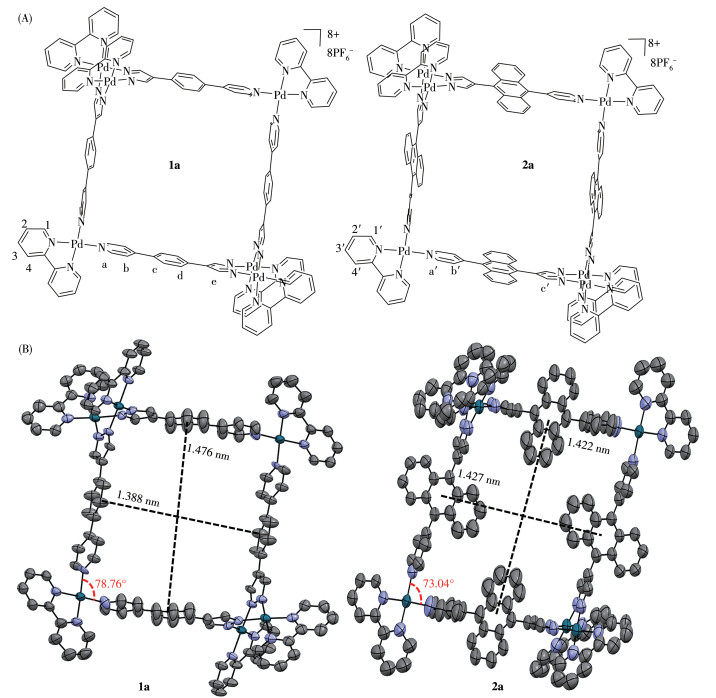
[Pd2(bpy)2 L2)2] (NO 3)2 and complex 2 were synthesised using an identical method. NMR analysis and mass spectroscopy confirmed all the ligands and primary assembly (Fig. S1-S9, S12-S15). Complexes 1a and 2a in the [4+4] mode were obtained by treating complexes 1 and 2 with KPF6, respectively. Fig. 1A shows the chemical structures of metallacycles 1a and 2a. The structures of the two metallacycles were further confirmed by singlecrystal X-ray diffraction (SCXRD) (Fig. 1B), nuclear magnetic resonance analysis (Fig. S10, S11, S16, S17) and element analysis. X-ray-quality single crystals of metallacycles 1a and 2a were acquired by dichloromethane or tetrahydrofuran vapour diffusion into CH3CN solution at room temperature. As illustrated in Fig. 1B, the single crystals of 1a and 2a exhibit parallelogram spatial configurations with acute angles of 78.76° and 73.04°, respectively. Single-crystal structures reveal cavity dimensions of 1.388 nm×1.476 nm for 1a and 1.427 nm×1.422 nm for 2a. Pd ions are located at the four corners of the metallacycle framework. The different lengths of Pd parallelograms may be attributed to the steric hindrance of the anthracene group in the supramolecular parallelogram. In addition, SCXRD analysis revealed that supramolecular Pd parallelograms 1a and 2a crystallize in the P1 and C2/m space groups, respectively (Table S1). The 1H NMR spectrum of 1a exhibited noteworthy downfield shifts for Ha (δ =8.62-9.28), Hb (δ =7.75-8.28), Hc (δ =7.73-7.85), Hd (δ=7.82-8.35) and He (δ=8.17-8.65) compared with the free ligand HL1 (Fig. S18). Similarly, the 1H NMR spectrum of 2a also exhibited downfield chemical shifts (Fig.S19). This result is attributed to the electron drawing effect of Pd2+, which is coordinated with the pyrazole units. The TGA curves demonstrated that 1a and 2a possess a large amount of solvent molecules in the crystal (Fig.S20). These results demonstrate that two supramolecular Pd parallelograms 1a and 2a are obtained through hierarchical self-assembly.
Figure 1
Figure 1.
Chemical structures (A) and single-crystal structures (B) of secondary assembly complexes 1a and 2a
All hydrogen atoms and hexafluorophosphate ions are omitted for clarity; Due to the presence of disordered structures of the anthracene ring, only one structure is present in the 2a crystal; The ellipsoid contour is at the 30%probability level.
Fig. 2 and 3 show the molecular packing of supramolecular Pd parallelograms 1a and 2a,respectively. As shown in Fig. 2, molecular stacking along the a- and c-axes through noncovalent interactions between hydrogen hexafluorophosphate anions and metallacycles. Conversely, C—H··· π interactions with 0.287 nm occur in the complex 1a crystal along the b-axis (Fig. 2B). Similarly, molecular packing along the a-, b-and c-axes through C—H···F interactions between hydrogen hexafluorophosphate anions and metallacy-cles also occur in the 2a crystal (Fig. 3). However, C— H··· π interactions with 0.285 nm along the a-axis (Fig. 3A) and C—H···H interactions with 0.245 nm along the c-axis (Fig. 3C) occur in the supramolecular Pd parallelogram complex 2a crystal. These data imply that the hydrogen hexafluorophosphate anion plays a crucial role in the growth of these supramolecular Pd parallelogram crystals.
Figure 2
Figure 2.
Molecular packing of complex 1a along the a-axis (A), b-axis (B) and c-axis (C) in the single-crystal state
Fig. S20 shows that the ligands and supramolecular Pd parallelograms had similar absorption peaks because they have the same aromatic structures in the supramolecular assembly. The absorption peak at 310 nm is the characteristic absorption peak of benzene. It was redshifted upon adding HSO3- to complex 1a. In complex 2a, tailing absorption bands at 360, 379 and 400 nm appeared due to the fingerprint peaks of the anthracene units (Fig. S21B). Upon adding HSO3- to complex 2a, the absorption bands were blueshifted. The fluorescence response of supramolecular Pd parallelograms on HSO 3- was investigated in DMSO-H2O mixtures with 50% water fraction at an excitation wavelength of 320 nm (Fig. 4A and 4B). Fig. 4A and 4B reveal that fluorescence was enhanced upon adding HSO3- in the 0-100 µmol·L-1 concentration range. Unlike the fluorescence titration spectra of complex 2a, the fluorescence spectra of complex 1a exhibited a shoulder peak at 500 nm upon adding HSO3- (Fig. 4A). Thus, the fluorescence of secondary assembly 1a in filter paper was turquoise after titration of HSO3-(Fig. 4E). However, after treating the strip with different anion solutions, the fluorescence of the other anions did not change under 365 nm ultraviolet excitation. In addition, the fluorescence intensity was linearly related to HSO3- concentration in a range of 30-80 µmol·L-1, indicating that complex 1a is suitable for quantitatively detecting HSO3- ions. The detection limit was calculated using the formula 3σ/k, where σ is the standard deviation of ten blank samples and k is the slope of the linear fitting plot. It was 0.131 µmol·L-1 in a mixture of DMSO and water with a water fraction of 50% (Fig. S22). We also investigated the fluorescence response of complex 1a at concentrations of 10 µmol·L-1 to the other anions (20 times the amount of the anion). Fig. 4C shows that bisulphite exhibited the most significant flu-orescence enhancement among the common anions. The I- and NO2- species also showed fluorescence enhancement in a range of 350-600 nm, but the intensity of the enhanced fluorescence emission caused by I- and NO 2- was only a quarter of that of HSO 3-. The fluorescence of the other 12 anions did not change significantly (Fig. 4C). Fig. 4D shows that several potential interfering substances, such as CN-, CO32-, Cl-, SO42-, SO32-, NO2-, NO3-, HPO42-, H2PO4-, F-, PO42-, Br-, I-, HSO 4-, did not affect the fluorescence response of 1a, thus confirming that the fluorescence sensor detects HSO 3- in water with high selectivity even in the pres-ence of competing anions. Similarly, the complex -2a- based sensor was robust against interference in aque-ous solution (Fig.S23).
Figure 4
Figure 4.
Fluorescence spectra of complexes 1a (A) and 2a (B) sensors with increasing HSO3- concentration; (C) Selectivity of 1a in DMSO-H2O mixtures with a water fraction of 50% towards HSO3- and other anions (c=10 µmol·L-1); (D)Photoluminescence intensity of 1a in a solution of DMSO-H2O mixtures treated with different anions (red) and the anions with HSO3- (yellow); (E) Fluorescence of 1a in test strip treated with different anions and illuminated by an ultraviolet lamp (365 nm)
To further investigate the detection mechanism of HSO 3-, 1H NMR titration experiments were conducted. The proposed reaction mechanism is illustrated in Fig. 5A and S24. When excessive HSO3- salt was added to the DMSO-d6 solution of the metallacycles, a new signal peak was generated, reminiscent of the free ligand HL1 region. This result indicates that metallacycles can dissociate and release free ligands (Fig. 5B).The 1 H NMR spectra also indicate that ligand HL1 binds a proton, resulting in an obvious fluorescence enhancement due to suppressed photoinduced electron transfer. In addition, HSO3- is a better receptor for [Pd2(bpy)2(NO3)2] (NO3)2, leading to the formation of Pd(bpy)(SO3)2[28].
Figure 5
Figure 5.
(A) Proposed disassembly mechanism of complex 1a with HSO3-; (B) Partial 1H NMR spectra (400 MHz, DMSO-d6) of ligands HL1, [Pd2(bpy)2(NO3)2](NO3) 2, complex 1a and bipyridine upon the addition of excess of HSO3-
In summary, two metal-organic supramolecular metallacycles based on dipalladium corners were synthesised. SCXRD shows that these metallacycles contain aryl pyrazole pyridine as linkers and Pd metal vectors as corners. Metallacycles 1a and 2a can be used as fluorescence turn-on sensors for detecting HSO3- with a low detection limit of 0.131 µmol·L-1. More important-ly, these metal complexes can selectively and sensitively detect HSO3- without interference from other ions.1H NMR titration experiments prove that upon adding HSO3-, the metallacycles can dissociate and release free ligands. The findings provide new insights into the development of metal-organic supramolecular sensors that exploit the stimulus-response destruction mechanisms of these metallacycles.
Supporting information is available at http://www.wjhxxb.cn
[1]
Sun Y Q, Liu J, Zhang J Y, Yang T, Guo W. Fluorescent probe for biological gas SO2 derivatives bisulfite and sulfite[J]. Chem. Commun.,
2013, 49(26):
2637-2639.
doi: 10.1039/c3cc39161b
[2]
朱根逸. 大气中二氧化硫监测技术的进展[J]. 分析化学,
1976(3): 224-234.
ZHU G Y. Progress in monitoring technology for sulfur dioxide in the atmosphere[J]. Chin. J. Anal Chem.,
1976, (3):
224-234.
[3]
Wang K N, Zhu Y L, Xing M M, Cao D X, Guan R F, Zhao S F, Liu Z Q, Mao Z W. Two-photon fluorescence probes for mitochondria imaging and detection of sulfite/bisulfite in living cells[J]. Sens. Actuator BChem.,
2019, 295:
215-222.
doi: 10.1016/j.snb.2019.05.077
[4]
Meng Z Q, Yang Z H, Li J L, Zhang Q X. The vasorelaxant effect and its mechanisms of sodium bisulfite as a sulfur dioxide donor[J]. Chemosphere,
2012, 89(5):
579-584.
doi: 10.1016/j.chemosphere.2012.05.056
[5]
Chan J, Dodani S C, Chang C J. Reaction-based small-molecule fluorescent probes for chemoselective bioimaging[J]. Nat. Chem.,
2012, 4:
973-984.
doi: 10.1038/nchem.1500
[6]
Li J, Gao Y, Guo H R, Li X K, Tang H Y, Li J, Guo Y. A novel colorimetric and ratiometric fluorescent probe for selective detection of bisulfite in real samples and living cells[J]. Dyes Pigment,
2019, 263:
285-290.
[7]
Deng C C, Xu Z Y, Sun Z, Xie J H, Luo H Q, Li N B. One-step synthesis of aldehyde-functionalized dual-emissive carbon dots for ratiometric fluorescence detection of bisulfite in food samples[J]. Food Chem,
2023, 405:
134961.
doi: 10.1016/j.foodchem.2022.134961
[8]
Chao J B, Wang H J, Zhang Y B, Yin C X, Huo F J, Sun J Y, Zhao M G. A novel pyrene-based dual multifunctional fluorescent probe for differential sensing of pH and HSO3- and their bioimaging in live cells[J]. New. J. Chem,
2018, 42(5):
3322-3333.
doi: 10.1039/C7NJ03903D
[9]
Guo L E, Zhang J F, Liu X Y, Zhang L M, Zhang H L, Chen J H, Xie X G, Zhou Y, Luo K, Yoon J. Phosphate ion targeted colorimetric and fluorescent probe and its use to monitor endogeneous phosphate ion in a hemichannel-closed cell[J]. Anal. Chem.,
2015, 87:
1196-1201.
doi: 10.1021/ac503818p
[10]
Zhang H Y, Huang Z J, Feng G Q. Colorimetric and ratiometric fluorescent detection of bisulfite by a new HBT-hemicyanine hybrid[J]. Anal. Chim. Acta,
2016, 920:
72-79.
doi: 10.1016/j.aca.2016.03.027
[11]
Wu W L, Ma H L, Huang M F, Miao J Y, Zhao B X. Mitochondriatargeted ratiometric fluorescent probe based on FRET for bisulfite[J]. Sens. Actuator B-Chem,
2017, 241:
239-244.
doi: 10.1016/j.snb.2016.10.028
[12]
Liu Y, Wu L Q, Dai Y X, Li Y P, Qi S L, Du J S, Yang Q B, Xu H, Li Y X. A novel fluorescent probe based on a triphenylamine derivative for the detection of HSO 3with high sensitivity and selectivity[J]. Anal. Methods,
2021, 13(33):
3667-3675.
doi: 10.1039/D1AY00800E
[13]
李芳, 唐永和, 郭锐, 林伟英. 高灵敏线粒体靶向近红外二氧化硫荧光探针的开发及细胞、小鼠成像研究[J]. 有机化学,
2021,41,(3): 1108-1116.
LI F, TANG Y H, GUO R, LIN W Y. Development of highly sensitive mitochondrial targeted nearinfrared sulfur dioxide fluorescent probes and imaging studies in cells and mice[J]. Chin. J. Org Chem,
2021, 41(3):
1108-1116.
[14]
Shi Q, Shen L Y, Xu H, Wang Z Y, Yang X J, Huang Y L, Redshaw C, Zhang Q L. A 1-hydroxy-2, 4-diformylnaphthalene-based fluorescent probe and its detection of sulfites/bisulfite[J]. Molecules,
2021, 26:
3064.
doi: 10.3390/molecules26113064
[15]
Tamima U, Singha S, Kim H R, Reo Y J, Jun Y W, Das A, Ahn K H. A benzocoumarin based two-photon fluorescent probe for ratiometric detection of bisulfite[J]. Sens. Actuator B-Chem,
2018, 277:
576-583.
doi: 10.1016/j.snb.2018.09.052
[16]
Han Y Y, Huang Y, Lin Q W, Tang L Y, Yang G Y, Xin H T, Zhao S F, Guan R F, Wang K N, Cao D X. Bifunctional fluorescent probe for the recognition of hydrazine and bisulfite in lipid droplets[J]. Sens. Actuator B-Chem.,
2023, 393:
134181.
doi: 10.1016/j.snb.2023.134181
[17]
Wang H, Wu X M, Yang S X, Tian H Y, Liu Y G, Sun B G. A dualsite fluorescent probe for separate detection of hydrogen sulfide and bisulfite[J]. Dyes Pigment.,
2019, 160:
757-764.
doi: 10.1016/j.dyepig.2018.09.020
[18]
Yang W C, Li S Y, Ni S N, Liu G Z. Advances in FRET-based biosensors from donor-acceptor design to applications[J]. Aggregate,
2023, 5(2):
e460.
[19]
王成, 杨曼, 邓祥义. 一种检测亚硫酸氢根的苯并咪唑类荧光增强型探针及其实际应用[J]. 无机化学学报,
2020,36,(4): 762-768.
WANG C, YANG M, DENG X Y. A benzimidazole based fluorescence enhanced probe for detecting bisulfite and its practical application[J]. Chinese J. Inorg. Chem,
2020, 36(4):
762-768.
[20]
Ma L, Haynes C J E, Grommet A B, Walczak A, Parkins C C, Doherty C M, Longley L, Tron A, Stefankiewicz A R, Bennett T D, Nitschke J R. Coordination cages as permanently porous ionic liquids[J]. Nat. Chem.,
2020, 12:
270-275.
doi: 10.1038/s41557-020-0419-2
[21]
Li Y C, Li X Z, Li L L, Xiao B, Wu J G, Li H C, Li D Y, He C. Phenoxazine-based supramolecular tetrahedron as biomimetic lectin for glucosamine recognition[J]. Chin. Chem. Lett.,
2021, 32(2):
735-739.
doi: 10.1016/j.cclet.2020.07.028
[22]
Woods C Z, Wu H T, Ngai C, Camara B D, Julian R R, Hooley R J. Modifying the internal substituents of self-assembled cages controls their molecular recognition and optical properties[J]. Dalton Trans.,
2022, 51:
10920-10929.
doi: 10.1039/D2DT01451C
[23]
Begato F, Licini G, Zonta C. Programmed guest confinement via hierarchical cage to cage transformations[J]. Chem. Sci.,
2023, 14(30):
8147-8151.
doi: 10.1039/D3SC01368E
[24]
Zhang D W, Ronson T K, Zou Y Q, Nitschke J R. Metal-organic cages for molecular separations[J]. Nat. Rev. Chem.,
2021, 5:
168-182.
doi: 10.1038/s41570-020-00246-1
[25]
Hou Y J, Wu K, Wei Z W, Li K, Lu Y L, Zhu C Y, Wang J S, Pan M, Jiang J J, Li G Q, Su C Y. Design and enantioresolution of homochiral Fe(Ⅱ)-Pd(Ⅱ) coordination cages from stereolabile metalloligands: Stereochemical stability and enantioselective separation[J]. J. Am. Chem. Soc.,
2018, 140:
18183-18191.
doi: 10.1021/jacs.8b11152
[26]
Kou Y L, Tong J, Meng C, Yuan Q, Wang J, Yu S Y. Reversible and turn-on fluorescence detection of phosphate in aqueous solution and living cell imaging by supramolecular metallacycles with AIE-active ligands[J]. ACS Appl. Mater. Interfaces,
2023, 15:
10828-40838.
[27]
Wang P P, Tong J, Meng C, Yuan Q, Deng W, Yu S Y, Ma H W. Self-assembly of tripyrazolate-linked[M6L2] cages for the selective sensing of HSO3- and gaseous SO2 by turn-on fluorescence[J]. Dalton Trans.,
2023, 52(19):
6129-6137.
[28]
Cui Y, Chen Z M, Jiang X F, Tong J, Yu S Y. Self-assembly and anion sensing of metal-organic[M6L2] cages from fluorescent triphe-nylamine tri-pyrazoles with dipalladium (Ⅱ, Ⅱ) corners[J]. Dalton Trans.,
2017, 46(18):
5801-5805.
doi: 10.1039/C7DT00179G
[29]
Liu Y, Liu F Z, Yan K K. Mechanochemical access to a short-lived cyclic dimer Pd2L2: An elusive kinetic species en route to molecular triangle Pd3L3 and molecular square Pd4L4[J]. Angew. Chem. Int. Ed.,
2022, 61(18):
e202116980.
doi: 10.1002/anie.202116980
[30]
Krishnaswamy S, Prusty S, Chartrand D, Hanan G S, Chand D K. Self-assembled molecular squares as supramolecular tectons[J]. Cryst. Growth Des.,
2018, 18:
2016-2030.
doi: 10.1021/acs.cgd.7b01425
[31]
Behnia A, Boyle P D, Fard M A. Pincer-plus-one ligands in self-assembly with palladium(Ⅱ): A molecular square and a molecular tetrahedron[J]. Dalton Trans.,
2016, 45(48):
19485-19490.
doi: 10.1039/C6DT04264C
[32]
Zhou M Y, Yu Z S, Deng W, Lu H L, Niu X F, Tong J, Yu S Y. [M8L4]8+-type squares self-assembled by dipalladium corners and bridging aromatic dipyrazole ligands for iodine capture[J]. Inorg Chem.,
2023, 62:
10193-10202.
doi: 10.1021/acs.inorgchem.3c00893
[33]
Gupta G, You Y J, Hadiputra R, Jung J, Kang D K, Lee C Y. Heterome-tallic BODIPY-based molecular squares obtained by self-assembly: Synthesis and biological activities[J]. ACS Omega,
2019, 4:
13200-13208.
doi: 10.1021/acsomega.9b01328
Figure 1
Chemical structures (A) and single-crystal structures (B) of secondary assembly complexes 1a and 2a
All hydrogen atoms and hexafluorophosphate ions are omitted for clarity; Due to the presence of disordered structures of the anthracene ring, only one structure is present in the 2a crystal; The ellipsoid contour is at the 30%probability level.
Figure 4
Fluorescence spectra of complexes 1a (A) and 2a (B) sensors with increasing HSO3- concentration; (C) Selectivity of 1a in DMSO-H2O mixtures with a water fraction of 50% towards HSO3- and other anions (c=10 µmol·L-1); (D)Photoluminescence intensity of 1a in a solution of DMSO-H2O mixtures treated with different anions (red) and the anions with HSO3- (yellow); (E) Fluorescence of 1a in test strip treated with different anions and illuminated by an ultraviolet lamp (365 nm)
Figure 5
(A) Proposed disassembly mechanism of complex 1a with HSO3-; (B) Partial 1H NMR spectra (400 MHz, DMSO-d6) of ligands HL1, [Pd2(bpy)2(NO3)2](NO3) 2, complex 1a and bipyridine upon the addition of excess of HSO3-

 下载:
下载:


 下载:
下载:


 下载:
下载:
