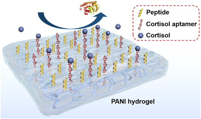
Scheme 1.
Schematic illustration of the antifouling biosensor.
A wearable electrochemical biosensor based on antifouling and conducting polyaniline hydrogel for cortisol detection in sweat
Xiujuan Qiao , Zhenying Xu , Zhen Wei , Yiting Hou , Fengxian Gao , Xijuan Yu , Xiliang Luo

The pace of life in the 21st century is undergoing exponential acceleration, subjecting individuals to a series of stresses deriving from their social and personal spheres. These stresses are harmful to both mental [1] and physical [2] well-being. The means for stress diagnosis and management involve quantifying cortisol release [3]. Cortisol, as a medical stress biomarker from the adrenal glands [4], is released during periods of stress and anxiety [5,6]. It is closely related to multiple psychological functions, such as regulating glucose levels [7], carbohydrate metabolism [8], and blood pressure [9]. The enhancement of cortisol concentrations will be harmful to weight, immune functionality, inducing chronic illnesses [10].
The rapid advancement of wearable sweat sensors has aroused unique opportunity for the seamless, real-time collection of personalized physiological data, offering continuous, non-invasive monitoring of various biomarkers through sweat analysis [11-13]. The concentration of cortisol in sweat typically ranges between 8 ng/mL and 140 ng/mL [14], which makes sweat a valuable biological fluid for monitoring cortisol levels [15]. Therefore, the integration of electrochemical sensors with wearable electronics provides an attractive method for the noninvasive detection of cortisol, which can be widely used in monitoring human health and mood [16,17].
Despite many advantages presented by wearable sweat sensors [18], the issue of inevitable biofouling poses obstacles to sustain sensor utilization [19-21], impeding their further application, popularization, and commercial viability [22]. Similar to complex blood components, sweat comprises a serious of components, including metabolic byproducts, electrolytes, vitamins, amino acids, metal ions, exogenous compounds (such as alcohol, drugs, nicotine), shed skin cells, trace protein, and significant quantities of oil [23-27]. Accurate detection in biosensing encounters a formidable challenge: (Bio)molecules in complex biological fluids exhibit nonspecific adsorption onto the sensing surface [28]. This phenomenon leads to the generation of false signals and the diminishment in the sensitivity of the biosensing system [29].
Therefore, it is urgent to construct antifouling biosensors capable of withstanding biological contamination for wearable detection [30]. In recent years, various antifouling materials, including polyethylene glycol, oligoethylene glycol, zwitterionic polymers, and polypeptides, have shown remarkable antifouling capabilities [31-33]. To deal with this issue, antifouling materials are introduced and applied as coatings during the construction of electrochemical sensors. These materials possess robust hydrophilic nature, forming a hydration layer which endows them with the capability to resist unspecific adsorption [34,35]. Among them, peptide-based biomaterials have gained attention as effective anti-fouling materials for biosensor development due to their beneficial physicochemical characteristics, excellent biocompatibility, and strong hydration capabilities [31].
Here, we have developed an antifouling wearable biosensor capable of precise cortisol detection. This antifouling wearable sensor was designed based on PANI hydrogel and hydrophilic polypeptides. PANI hydrogel has its unique water storage property to resist unspecific adsorption. At the same time, the increased specific surface area due to PANI hydrogel's three-dimensional (3D) structure facilitated the attachment of numerous antifouling peptides, contributing to the formation of dense antifouling structure. This setting ensures the accuracy of target cortisol detection in complex sweat (Scheme 1). To validate the biosensor's efficacy, we conducted cortisol level measurements on volunteers during both morning and evening. The observed data followed distinct circadian rhythm, exhibiting similar results compared to commercial ELISA kit. Therefore, this electrochemical biosensor achieves successful monitoring for cortisol concentration in human sweat, providing potential for practical application among wearable area.
The experimental procedures were outlined in Supporting information.
For preparation of antifouling electrochemical biosensor, PANI hydrogel were firstly electrodeposited on the carbon screen-printed electrodes (CPE) surface. Scanning electron microscopy (SEM) results illustrated that PANI possessed a three-dimensional cross-linked hydrogel network structure with an average pore size of 100–200 nm (Figs. 1A and B). The differential pulse voltammetry (DPV) test was then used to characterize the antifouling sensor construction process in PBS (0.2 mol/L, pH 7.0) solution. As shown in Fig. S1A (Supporting information), there was no signal generation of bare CPE in PBS solution. After electrodeposition of PANI hydrogel, there was an obvious peak at −0.05 V, corresponding to the oxidation of PANI hydrogel, which proved the successful deposition of PANI hydrogel. When the non-conductive peptides and aptamers (APTA) were introduced, the current value decreased due to their decrease of charge transfer ability in the solution. Following the attachment of the target cortisol, the DPV signal further reduced because the specific recognition between the target and the aptamers increased the impedance and thereby reduced the electron transfer ability. Furthermore, as shown in Fig. S1B (Supporting information), the corresponding opposite trend of each modification process obtained by electrochemical impedance spectroscopy (EIS) also described the successful construction of this biosensor and the feasibility of cortisol detection.
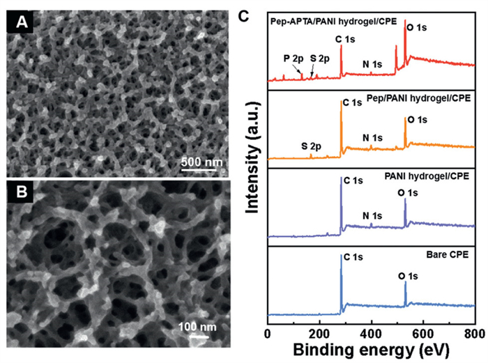
In addition, the biosensor preparation process was further characterized using X-ray photoelectron spectroscopy (XPS). The XPS technique is an important surface-sensitive quantitative spectroscopic technique that is frequently used to measure the elemental composition of the near surface region. The main information of XPS was obtained at the uppermost levels of the composition profile of prepared electrode process. As illustrated in Fig. 1C, the bare CPE primarily was consisted of carbon (C) and oxygen (O) elements. Following the electrodeposition of the PANI hydrogel, a distinct peak corresponding to nitrogen (N) could be clearly observed. Upon subsequent binding of the peptide, a characteristic sulfur (S) peak appeared, accompanied by a slight increase in the N 1s peak. This observation demonstrated the successful binding of the peptide. Additionally, as the aptamer contains a phosphate backbone with phosphorus (P), the characteristic peak of P was evident when the aptamers were immobilized onto the electrode. These XPS characterization results collectively indicated the successful construction of the biosensor.
To establish the most effective antifouling sensing platform, it is essential to optimize the conditions for antifouling and sensing. This can be achieved by investigating the impact of various factors on sensing performance, including the effect of incubation time of peptides and aptamers, the aptamer's concentration, as well as the connection time of the target cortisol.
By incubating aptamers with varying concentrations on the electrode surface, the effect of aptamer concentration was investigated, as illustrated in Fig. S2A (Supporting information). Changes in DPV currents were recorded before and after incubation. The point at which the signal suppression rate (%) essentially reached equilibrium for 1.5 h indicated the saturation of aptamer incubation concentration (1 µmol/L). The results demonstrated that the signal suppression rate gradually rose as the aptamer concentration increased. The optimal concentration of aptamer was identified as 1 µmol/L. The optimization of peptide and aptamer cultivation time was conducted with the peptide of 2 µmol/L and the aptamer of 1 µmol/L. As depicted in Fig. S2B (Supporting information), the binding of the peptide and aptamer to the substrate reached saturation after 1.5 h. Consequently, the optimal cultivation time was determined to be 1.5 h. And Fig. S2C (Supporting information) clearly illustrated that the signal suppression rate reached its maximum at a connection time of 45 min, indicating equilibrium between the aptamer and the target cortisol molecule. Therefore, the optimal cortisol connection time was determined to be 45 min.
Wearable sensors designed for direct application in sweat detection must exhibit robust resistance to non-specific adsorption of contaminants in sweat. To assess the non-specific adsorption ability of the biosensor during actual detection, DPV technique was conducted to investigate signal changes for long time in various modified interfaces after immersing in artificial sweat, including bare CPE (Fig. 2A), PANI film/CPE (Fig. 2B), PANI hydrogel/CPE (Fig. 2C), and Pep/PANI hydrogel/CPE (Fig. 2D). Every cultivating time is 30 min in 100% artificial sweat. Concurrently, statistical results were presented in Fig. 3.
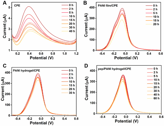
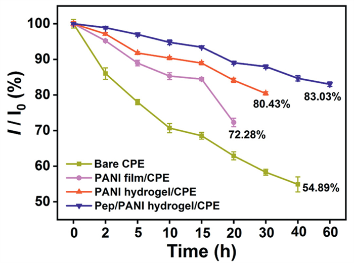
The signal of bare electrode changed most obviously with the original signal decreasing to 54.89% after soaking for 40 h. The signal of PANI membrane electrode decreased sharply after 15 h, and decreased to 72.28% after 20 h. However, the signal of PANI hydrogel modified electrode remained at 80.43% after soaking in artificial sweat for 30 h, which can be explained by the hydrogel's excellent three-dimensional (3D) network structure. This structure endowed the electrode with good water storage property, and thus forming a hydrophilic layer to resist the adsorption of pollutants. Furthermore, upon introducing the antifouling polypeptide, the hydrogel surface formed a dense antifouling layer. This layer acted as a potent defense against fouling blocking the electrode surface, ensuring that the signal sustained at 83.03% of its initial strength even after 60 h These results provide evidence for biosensor's potential suitability in long-term sweat monitoring as a wearable device.
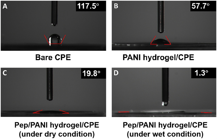
The hydrophilicity is a key indicator for the evaluation of electrode's antifouling performances. As depicted in Fig. 4, the water contact angle of the PANI hydrogel electrode decreased to 57.7° in comparison to the unmodified electrode. After the attachment of the polypeptide, the water contact angle further diminished to 19.8° in the dry state and decreased to 1.3° in the wet state. This could be attributed to hydrogel's robust water storage capability. This proved the strong hydrophilicity of the sensing interface based on antifouling and conducting polyaniline hydrogel.
To provide a visual representation of the sensor's antifouling capability, we assessed the adsorption extent of fluorescein isothiocyanate (FITC-BSA) using laser confocal imaging. Bare ITO, PANI film/ITO, PANI hydrogel/ITO, and Pep/PANI hydrogel/ITO were individually immersed in 0.2 mg/mL FITC-BSA solution for 30 min. As depicted in Fig. S3 (Supporting information), both bare CPE and PANI film/ITO exhibited robust green fluorescence, indicating significant FITC-BSA adsorption. However, the fluorescence intensity gradually decreased on PANI hydrogel/ITO and Pep/PANI hydrogel/ITO. This observation proved that the combination of PANI hydrogel and the antifouling polypeptide effectively enhanced the electrode's resistance to protein adsorption.
Upon the sensor's optimal conditions, cortisol detection was conducted. We employed DPV technology to measure the electrochemical current response of the sensor in PBS solution (0.2 mol/L, pH 7.0). As illustrated in Fig. 5A, the DPV signal of PANI decreased as the concentration of target cortisol increased. This phenomenon could be attributed to the specific binding of cortisol to the aptamer, which impeded the electron transfer process of PANI. Fig. 5B revealed that the signal suppression rate exhibited a robust linear correlation with the logarithm of concentration (Lg C, 10–10 - 10–6 g/mL), with an impressive linear correlation coefficient (R2 = 0.9989). Notably, the biosensor demonstrated a remarkable minimum detection limit for cortisol, capable of detecting concentrations as low as 33 pg/mL (S/N = 3).
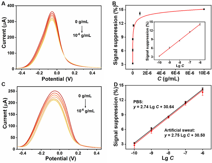
To assess the antifouling electrochemical biosensor's applicability for wearable detection, we conducted experiments to evaluate its biosensing response to the target molecule cortisol in artificial sweat (Fig. 5C). As depicted in Fig. 5D, the linear fit curve of the calculated signal suppression rate (red line) was similar to that in the buffer solution (Fig. 5D, black line). Notably, while the signal peaks in artificial sweat differed from those in the PBS buffer solution, the biosensor exhibited consistent sensing performance even in the presence of complex artificial sweat. This illustrated the potential feasibility of utilizing this antifouling electrochemical biosensor for practical application.
To further assess the selectivity of the biosensor, we examined the recognition of interfering substances commonly found in sweat by the sensing interface (Fig. 6A). The results demonstrated that the sensor exhibited substantial signal response to the target cortisol. Even in the presence of other sweat components such as steroid hormones (corticosterone, cortisone), adrenaline, uric acid (UA), ascorbic acid (AA), and lactic acid (LA), the signal suppression rate of the interface towards these interfaces is only mere 0.79%, indicating that the biosensor has excellent selectivity.
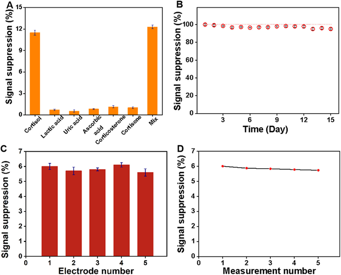
To evaluate the long-term stability of the sensors, three independently prepared biosensors were stored in a PBS solution up to 15 days. As depicted in Fig. 6B, the sensor still maintained a baseline signal of > 95%. To further investigate the reproducibility of the biosensor, 1.0 ng/mL cortisol was first measured using five independently prepared biosensors with a relative standard deviation (RSD) of 3.4% (Fig. 6C). At the same time, the same biosensor was used to measure the DPV signal of 1.0 ng/mL cortisol for 5 times at an interval of 30 min, and the RSD was 1.71% (Fig. 6D). The above results all proved that the biosensor has excellent reproducibility.
Previous research has verified that cortisol concentrations in sweat are close to those in the blood, exhibiting a day-night cycle characterized by higher levels in the morning (AM) and lower levels in the afternoon (PM) [36]. To further elucidate the relationship between sweat and circulating cortisol levels, we conducted an analysis of morning and afternoon sweat samples collected from four healthy subjects.
Before practical applications, the biosensor was placed on the skin's surface and undergone deformation as the body's joints move. To assess the sensor's deformation stability, a comprehensive evaluation was conducted under the condition of outward and inward bending, as well as stretching deformation. As demonstrated in Fig. 7A, the sensor's signal remained largely unaffected under a series of deformation tests. These results affirmed that the sensor exhibited exceptional mechanical stability, rendering it suitability for wearer's the daily activity. In addition, the base material of wearable sensor showed the better rebound resilience (Fig. S4 in Supporting information), excellent tensile property, and did not have irritative anaphylaxis (Fig. S5 in Supporting information).
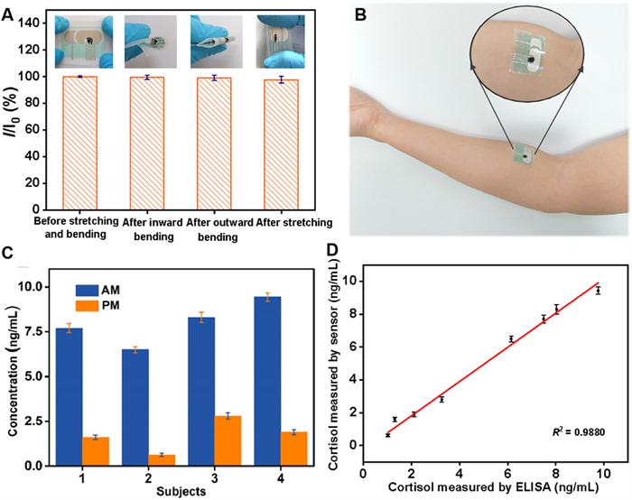
We then utilized a wearable patch to measure cortisol concentrations on arm (Fig. 7B). As illustrated in Fig. 7C, a consistent trend of cortisol regulation by circadian rhythm was evident across all samples. Additionally, we compared the results obtained using the sensor with those derived from commercial ELISA kits, yielding a remarkable correlation coefficient of R2 = 0.9880 (Fig. 7D). These above results showed the sensor's potential for integration into flexible wearable devices for cortisol detection.
In summary, we have successfully developed a wearable antifouling electrochemical sensor for cortisol detection. The incorporation of PANI hydrogel with the immobilization of peptides enhanced the precision of cortisol detection within the complex sweat environment. The biosensor has demonstrated its ability to accurately detect cortisol in both buffer solution and artificial sweat, covering a detection concentration range from 10−10 g/mL to 10−6 g/mL. Impressively, it boasts an exceptional minimum detection limit of 33 pg/mL, while maintains robust stability, outstanding selectivity, and reproducibility. Notably, this antifouling biosensor can be seamlessly integrated into wearable devices for the monitoring of cortisol circadian rhythms in sweat. This research holds significant promise in the field of personal medicine and the continuous health monitoring of individuals.
The sweat samples were provided from adult volunteers from our laboratory, as well as the informed consent for use of the human sweat was obtained. All sample preparations were approved by the relevant Institutional Review Committee and carried out in accordance with institutional guidelines and conformed to the relevant regulatory standards Ethical.
The authors declare that they have no known competing financial interests or personal relationships that could have appeared to influence the work reported in this paper.
Xiujuan Qiao: Writing – original draft, Methodology, Conceptualization, Funding acquisition. Zhenying Xu: Software, Methodology, Investigation, Data curation. Zhen Wei: Visualization, Investigation. Yiting Hou: Formal analysis, Data curation. Fengxian Gao: Writing – review & editing, Supervision, Funding acquisition. Xijuan Yu: Writing – review & editing, Validation. Xiliang Luo: Writing – review & editing, Supervision, Funding acquisition.
This work was supported by the National Natural Science Foundation of China (Nos. 22174082, 22374085, 22105113), the Key Research and Development Program of Shandong Province (No. 2021ZDSYS30), and Natural Science Foundation of Shandong Province, China (No. ZR2024QB059).
Supplementary material associated with this article can be found, in the online version, at doi:
A. Zivony, Nat. Hum. Behav. 3 (2019) 1037-1037. doi: 10.1038/s41562-019-0735-y
M. Kivimäki, A. Bartolomucci, I. Kawachi, Nat. Rev. Endocrinol. 19 (2023) 10–27. doi: 10.1038/s41574-022-00746-8
G. Russell, S. Lightman, Nat. Rev. Endocrinol. 15 (2019) 525–534. doi: 10.1038/s41574-019-0228-0
E. Dziurkowska, M. Wesolowski, J. Clin. Med. 10 (2021) 5204. doi: 10.3390/jcm10215204
A.J. Steckl, P. Ray, ACS Sens. 3 (2018) 2025–2044. doi: 10.1021/acssensors.8b00726
E. Mansour, W. Saliba, Y.Y. Broza, et al., ACS Sens. 8 (2023) 3215–3224. doi: 10.1021/acssensors.3c00945
N.K. Singh, S. Chung, M. Sveiven, et al., ACS Omega 6 (2021) 27888–27897. doi: 10.1021/acsomega.1c03552
J.E. An, K.H. Kim, S.J. Park, et al., ACS Sens. 7 (2022) 99–108. doi: 10.1021/acssensors.1c01734
M. Kivimäki, A. Steptoe, Nat. Rev. Cardiol. 15 (2018) 215–229. doi: 10.1038/nrcardio.2017.189
M.S. Sagmeister, L. Harper, R.S. Hardy, Front. Endocrinol. 13 (2023) 1075809. doi: 10.3389/fendo.2022.1075809
K. Di, J. Wei, L. Ding, et al., Chin. Chem. Lett. 36 (2025) 109911. doi: 10.1016/j.cclet.2024.109911
L. Xu, X. Hu, S. Zhou, et al., Chin. Chem. Lett. 35 (2024) 109103. doi: 10.1016/j.cclet.2023.109103
J. Min, J. Tu, C. Xu, et al., Chem. Rev. 123 (2023) 5049–5138. doi: 10.1021/acs.chemrev.2c00823
X. Weng, Z. Fu, C. Zhang, et al., Anal. Chem. 94 (2022) 3526–3534. doi: 10.1021/acs.analchem.1c04508
S. Madhu, A.J. Anthuuvan, S. Ramasamy, et al., ACS Appl. Electron. Mater. 2 (2020) 499–509. doi: 10.1021/acsaelm.9b00730
T. Laochai, J. Yukird, N. Promphet, et al., Biosens. Bioelectron. 203 (2022) 114039. doi: 10.1016/j.bios.2022.114039
M. Zea, F.G. Bellagambi, H. Ben Halima, et al., TrAC-trend, Anal. Chem. 132 (2020) 116058.
M. Bariya, H.Y.Y. Nyein, A. Javey, Nat. Electron. 1 (2018) 160–171. doi: 10.1038/s41928-018-0043-y
X. Wang, A. He, B. Yu, et al., Anal. Chem. 94 (2022) 14402–14409. doi: 10.1021/acs.analchem.2c03158
K. Koren, C.M. McGraw, ACS Sens. 8 (2023) 2432–2439. doi: 10.1021/acssensors.3c00961
L. Tao, Y. Kong, Y. Xiang, et al., Chin. Chem. Lett. 34 (2023) 107481. doi: 10.1016/j.cclet.2022.04.079
X. She, X. Wang, P. Niu, et al., Chem. Eng. J. 431 (2022) 133354. doi: 10.1016/j.cej.2021.133354
K. Van Hoovels, X. Xuan, M. Cuartero, et al., ACS Sens. 6 (2021) 3496–3508. doi: 10.1021/acssensors.1c01403
D. Sim, M.C. Brothers, J.M. Slocik, et al., Adv. Sci. 9 (2022) 2104426. doi: 10.1002/advs.202104426
H. Zhao, X. Zhang, Y. Qin, et al., Adv. Funct. Mater. 33 (2023) 2212083. doi: 10.1002/adfm.202212083
S. Upasham, N.K.M. Churcher, P. Rice, et al., ACS Sens. 6 (2021) 659–672. doi: 10.1021/acssensors.0c02622
Z.Q. Wu, X.Q. Cao, Y. Hua, et al., Anal. Chem. 96 (2024) 3087–3095.
K. Jayakumar, A. Lielpetere, D.A. Domingo-Lopez, et al., Biosens. Bioelectron. 219 (2023) 114815. doi: 10.1016/j.bios.2022.114815
M.J. Russo, M. Han, P.E. Desroches, et al., ACS Sens. 6 (2021) 1482–1507. doi: 10.1021/acssensors.1c00390
X. Qiao, Y. Cai, Z. Kong, et al., ACS Sens. 8 (2023) 2834–2842. doi: 10.1021/acssensors.3c00778
X. Qiao, Z.H. Qian, W. Sun, et al., Anal. Chem. 95 (2023) 11091–11098. doi: 10.1021/acs.analchem.3c01843
S. Zhao, X. Qiao, M. Chen, et al., ACS Sens. 7 (2022) 1740–1746. doi: 10.1021/acssensors.2c00518
J. Li, L. Zhang, P. Huang, et al., Chin. Chem. Lett. 36 (2025) 109970. doi: 10.1016/j.cclet.2024.109970
C. Jiang, G. Wang, R. Hein, et al., Chem. Rev. 120 (2020) 3852–3889. doi: 10.1021/acs.chemrev.9b00739
Z. Song, R. Li, X. Yang, et al., Chin. Chem. Lett. 34 (2023) 108314. doi: 10.1016/j.cclet.2023.108314
C.F. Sharpley, K.G. Kauter, J.R. McFarlane, Clin. Med. Insights-En. 3 (2010) CMED.S4350, doi: 10.4137/cmed.s4350.

Figure 1 (A, B) SEM of the PANI hydrogel. (C) XPS spectra of the different modified electrodes.

Figure 2 The antifouling performances of bare CPE recorded in PBS containing 5.0 mmol/L [Fe(CN)6]3-/4- and 0.1 mol/L KCl for long time after incubation in artificial sweat for 30 min, respectively (A). The antifouling performances of PANI film/CPE (B), PANI hydrogel/CPE (C), and Pep/PANI hydrogel/CPE (D) recorded in PBS (0.2 mol/L, pH 7.0) for long time after incubation in artificial sweat for 30 min, respectively.

Figure 3 The signal suppression (%) of the different electrodes after incubation in artificial sweat for long time.

Figure 4 Water contact angle images of bare CPE (A), PANI hydrogel/CPE (B), Pep/PANI hydrogel/CPE under dry condition (C) and Pep/PANI hydrogel/CPE under wet condition (D).

Figure 5 (A) DPV responses of the biosensor to target cortisol in PBS (0.2 mol/L, pH 7.0, 0 g/mL - 10–6 g/mL). (B) Signal suppression rate of the biosensor after incubation with various concentrations of cortisol in PBS. Inset showed the corresponding calibration curves. (C) DPV responses of the biosensor to target cortisol in artificial sweat. (D) The corresponding calibration curves of the biosensor after incubation with various concentrations of cortisol in PBS (black line) and in artificial sweat (red line).

Figure 6 (A) Signal suppression rates of the biosensor in response to 100 ng/mL cortisol, and 500 ng/mL of LA, UA, AA, corticosterone, cortisone, as well as their mixture, respectively. (B) Stability of the fabricated biosensor after being stored in PBS (0.2 mol/L, pH 7.0) for 15 days. (C) Signal suppression rates observed with five distinct biosensors during the detection of 1.0 ng/mL cortisol. (D) Discontinuous detection of 1.0 ng/mL cortisol by a single electrode for 5 times at an interval of 30 min.

Figure 7 (A) Relative DPV responses after 10 such repeated bending tests in PBS (0.2 mol/L, pH 7.0) involving inward bending, outward bending and stretching. (B) The image of CPE on human arm. (C) Cortisol levels in sweat in the AM and PM from four healthy subjects. (D) Correlation of cortisol concentration measured by ELISA and sensor.

 扫一扫看文章

扫一扫关注我们
 DownLoad:

 下载:
下载:
 下载:
下载: