Scheme 1.
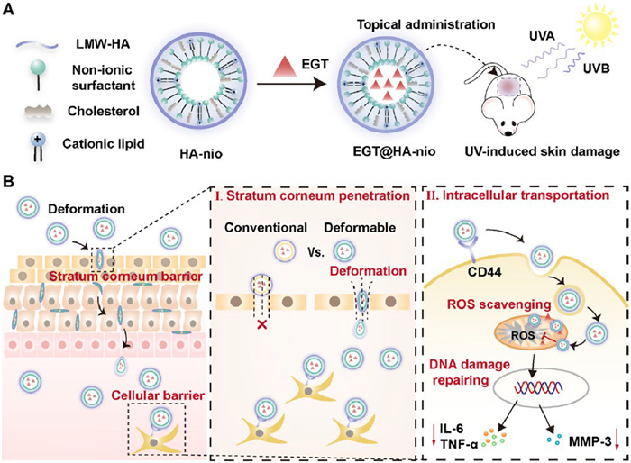
Schematic illustration of EGT@HA-nio preparation (A) and their therapeutic effect against UV-induced skin damage (B).
Deformable hyaluronic acid niosomes overcome multi-barriers for improved ergothioneine transdermal delivery against UV-induced skin damage
Lijun Li , Chenliang Guo , Yuelin Fang , Zijian Cheng , Yaowei Li , Zhangyu Wang , Dian Cai , Yuqi Xu , Wenqi Liu , Shouwei Ma , Xinxin Zhang

Transdermal administration remains a real challenge as it is difficult to penetrate the primary barrier of the skin, the stratum corneum (SC) [1]. Researchers have developed many strategies to improve the transdermal absorption of drugs, such as adding transdermal absorption enhancers [2], microneedles [3], and ultrasound [4]. However, these methods may irritate the skin or damage the SC to varying degrees, making them unsuitable for some skin diseases requiring noninvasive, modest, and long-term protection, like ultraviolet (UV)-induced skin damage. UV exposure triggers the generation and accumulation of reactive oxygen species (ROS), which further stimulates DNA damage and inflammatory response, leading to the development of skin aging and even carcinogenesis [5]. The transdermal delivery of drugs or active agents, especially antioxidants, has been promising for preventing UV-induced skin damage [6]. Various transdermal drug delivery systems (TDDS), such as liposomal formulations, microemulsions and hydrogel systems, have demonstrated their effectiveness in delivering antioxidants against UV-induced skin damage [7–10]. However, their delivery of drugs was reported to be limited to the upper layers of skin. TDDS are required to overcome the multiple physiological barriers for the ideal effect. Effective TDDS design requires strategies to penetrate the stratum corneum, traverse the epidermis, and achieve intracellular delivery to ensure the desired pharmaceutical effects.
The physicochemical properties of TDDS, such as size [11], shape [12], surface chemistry [13], and lipophilicity [14], have been optimized to affect the absorption of topically applied agents through the skin. For instance, the phospholipids in the liposomes could fuse with the lipid layer of the skin and play a role in reducing the skin barrier to promote transdermal delivery [15]. Solid lipid nanoparticles (SLN) and nanostructured lipid carriers (NLC) can form an occlusive film at the SC surface which supports active molecule retention [16]. However, common TDDS design usually pays insufficient attention to penetrating the skin in their complete form, limiting the subsequent intracellular drug delivery process. To prevent TDDS from rupturing when penetrating through the SC, regulating the rigidity of nanoparticles may provide a viable solution [17]. Satisfactory deformability can push vesicles through smaller pores than average diameter, which prevents the active compound from being a naked state after penetration and ensures the subsequent intracellular uptake unaffected [18]. Consequently, taking mechanical softness into consideration when designing nanotherapeutics is reasonable for deep penetration into various biological barriers.
Niosomes (nio) made up of non-ionic surfactants are novel promising TDDS with advantages over conventional liposomes [19]. Herein, this study provides a new solution for treating UV-induced skin damage by preparing hyaluronic acid (HA)-coated niosomes (HA-nio) to deliver ergothioneine (EGT) (EGT@HA-nio). EGT is an attractive mitochondrially targeted antioxidant with a specialized uptake mechanism through organic cation transporter 1 (OCTN-1) (Scheme 1) [20,21]. Steareth-2 (non-ionic surfactant), cholesterol, and cationic lipid were chosen to assemble deformable niosomes (nio). Single tail non-ionic surfactant can act as membrane-softening agent to increase the deformability of vesicle membranes [22]. With optimal ratio of appropriate lipids, nio may offer flattering properties for transdermal delivery purposes in transdermal efficiency [23,24]. Meanwhile, the modification of HA would enhance the delivery of EGT to the skin cells, resulting in improved EGT distribution in mitochondria without the restriction of OCTN-1 transporter. Overall, HA-nio are expected to produce superior anti-UV-induced skin damage efficacy through overcoming multiple skin biological barriers.
To demonstrate the advantages of deformable HA-nio compared with conventional TDDS, HA modified liposomes (HA-lip) were chosen as the positive control group. The composition of HA-nio was shown in Fig. 1A. Non-ionic surfactant steareth-2 (S2), cholesterol (chol) and cationic lipids were added to enhance the stability and delivery efficiency by regulating membrane integrity and rigidity, and HA was modified on the surface to achieve better intracellular delivery efficiency. The successful preparation of niosomes is mainly affected by the ratio of non-ionic surfactant to cholesterol. Thus, we evaluate the diameter and polymer dispersity index (PDI) of nanoparticles formulated by different ratios of non-ionic surfactant to chol to obtain the optimized formulation. As shown in Fig. 1B, when the ratio of cholesterol to non-ionic surfactant or phospholipid (phos) is 1:1, the mean diameter of three nano-formulations was approximately 150 nm and PDI was around 0.2, which reveals higher stability [25]. Unless otherwise noted, this proportion of formulations was used for the following experiments.

The morphology of blank nio, HA-lip, and HA-nio was first characterized by transmission electron microscope (TEM) (Fig. 1C). The images show that nio, HA-lip, and HA-nio had spherical morphology and uniform size distribution. The HA-lip and HA-nio images display that the outer layers of liposome and niosome were wrapped by the HA layer. The dynamic light scattering (DLS) results show that the average diameter of HA-nio was 176 nm, slightly larger than that of nio as a result of HA camouflage (Fig. 1D). HA modification changed the zeta potential from 20.2 mV (blank nio) to 14.0 mV (HA-nio) due to its negative charge, and HA-lip show a negative zeta potential around −20 mV (Fig. 1E). Theoretically, compared to negatively charged HA-lip, positively charged HA-nio and nio would have a more robust interaction with the negatively charged skin surface, which may facilitate nio and HA-nio to penetrate the skin. The encapsulation efficiency of EGT in nio, HA-lip, and HA-nio determined by high performance liquid chromatography (HPLC) was approximately equal, all over 90% (Fig. 1F). The approximately equal encapsulation efficiency of nio and HA-nio indicate that HA modification had no influence on the drug encapsulation. The sizes of nio, HA-lip and HA-nio remained stable over 7 days, suggesting the feasibility for long-term use (Fig. S1 in Supporting information). Additionally, to evaluate the cytotoxicity, the effect of different doses of formulations on human dermal cells (HDFs) survival rate was studied. The results show that within the tested concentration range (10–1000 µg/mL), the cell survival rate was rarely affected, suggesting the good cytotoxicity of these formulations (Fig. 1G).
For transdermal delivery, niosomal vesicles were reported to present better flexibility to overcome the SC barrier and physicochemical stability [19]. To explore the degrees of deformation under physiological stress, a scan in liquid probe in contact mode of atomic force microscopy (AFM) was applied to measure the mechanical stiffness of nanoparticles. The Young's modulus of both HA-nio and nio was approximately 10 MPa, which is 1.45-fold lower than that of the HA-lip (14.5 MPa), further confirming that they have higher mechanical flexibility (Fig. 1H). The morphologies of nio, HA-lip, and HA-nio were characterized using AFM-based nanomechanical mapping. As shown in Fig. 1C, nio, HA-lip and HA-nio exhibited spherical morphology. With the force increased from 300 pN to 1000 pN, nio and HA-nio gradually deformed into different shapes while HA-lip displayed negligible deformation (Figs. 1I–K), suggesting nio and HA-nio had better deformability. The deformability of the nanocarriers was further assessed by examining their force–displacement responses against a probe. In the resulting force–indentation curves, variations in deflection during the probe's extension and retraction process indicated deviations in surface slope of the tested particles, serving as an indicator of particle deformation. As shown in Fig. 1M, no notable deflection error occurred for HA-lip, suggesting constantness throughout the test. However, nio and HA-nio exhibited significant deflection shifts during retraction, suggesting droplet deformation under probe compression (Figs. 1L and N). Outstanding deformability may be beneficial for nio and HA-nio to squeeze through the circa 75 nm intercellular space between the SC cells without rupture.
The impressive deformability of HA-nio sparks our interest and curiosity in exploring its ability to penetrate the SC of the skin (Fig. 2A). Initially, Franz diffusion cell with a polycarbonate membrane, whose pore size is 100 nm close to the intercellular space [26], was used to simulate the permeation process of DiI/DiO-labeled nio, HA-lip or HA-nio passing through the SC. Fluorescence resonance energy transfer (FRET) efficiency of DiO and DiI was an indicator of the integrity of dual-labeled nano-formulations. As is shown in Fig. 2B, the fluorescence intensity at wavelength 570 nm increases sequentially in nio, HA-lip, and HA-nio group, suggesting that compared to nio or HA-lip, more HA-nio passed through the pore in the polycarbonate membrane with better integrity own to the favorable deformability of HA-nio, paving a way for the subsequently active molecule release.
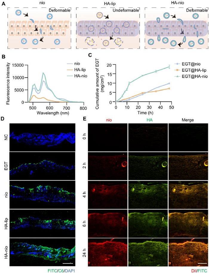
Next, we wondered whether HA-nio performed outstandingly in penetrating nude mice skin. Therefore, EGT release was determined on Franz diffusion cell by using the back skin of nude mice (Fig. 2C). The EGT@HA-lip released EGT faster than EGT@nio does at the early stage, possibly resulting from the hydration effect of HA. HA forms a hydration film in the SC, which can increase skin permeability, making it easier for active substances to penetrate the skin barrier and enter deeper layers of the skin [27]. However, at the later stage, the EGT@nio released EGT faster than EGT@HA-lip does, which may be ascribed to the deformability of EGT@nio starting to show a dominant role and making a large penetration amount of EGT@nio into the skin. Compared with EGT@HA-lip and EGT@nio, the EGT release of EGT@HA-nio was about 5-time higher at 12 h and maintained the top position during the recorded 50 h. As suggested by the highest FRET efficiency during the skin penetration evaluated before, HA-nio could pass through the intercellular space intactly benefiting from the deformability property, and the skin hydration effect of HA further enhanced the skin penetration, both contributing to the excellent transdermal drug delivery effect exhibited by HA-nio.
Then we investigated the distribution of EGT@nio, EGT@HA-nio, and EGT@HA-lip in vivo. All the animal experiments were conducted in compliance with protocols approved by Institutional Animal Care and Use Committee (IACUC) guidelines of the Shanghai Institute of Materia Medica (IACUC code: 2023–10-GY-71). BALB/c null mice were topically applied with fluorescein isothiocyanate (FITC)-EGT and coumarin 6 (C6)-labeled nano-formulations for 6 h and then cut the applied skin for further analysis. As shown in Fig. 2D, FITC accumulated outside the skin because of their hydrophilic property. Due to the deformability, nio can penetrate the skin but mainly accumulate in the SC layer. Owing to the skin hydration effect of HA, most HA-lip were distributed in SC layer and a few can penetrate to the dermis. Compared to nio and HA-lip, the fluorescence signal of the dermis layer in the HA-nio group was much stronger, demonstrating that more HA-coated noisome could pass through the SC to reach the dermis layer, consistent with the release behavior evaluation result. To further preliminarily predict whether HA-nio can exert potent effects in human skin, 2–3 months old pig skin with more similar physiological characteristics to those of humans was chosen as the transdermal absorption research model. According to the fluorescent microscopic results (Fig. 2E), the fluorescence of HA and nio overlapped, indicating that HA-nio could penetrate the skin in a completed form in line with the former results.
The above results demonstrated that the deformability and the skin hydration effect endow the HA-nio with superior permeability characteristic, which is conducive to the delivery of bioactive substances into the dermis cells.
As a potential mitochondrially targeted natural antioxidant, EGT cannot penetrate the cell membrane but requires a special transporter protein OCTN-1 to enter the cell, which limits its therapeutic effect [28]. Studies have shown that the CD44 receptor is highly expressed in HDFs and has a strong affinity for the HA molecule [29]. Coupled with the hydration of the skin by HA, the use of HA-nio for the delivery of EGT may significantly improve its cellular uptake.
To evaluate whether the enhanced cellular uptake capability of HA-nio meets our expectations, nio, HA-lip, and HA-nio were labeled with C6 for confocal laser scanning microscope (CLSM) imaging and flow cytometry (FCM) analysis (Figs. 3A–C). The results show that HA-nio-treated cells exhibited stronger C6 fluorescence intensity compared to cells treated with nio or HA-lip. This trend was confirmed by FCM quantitative analysis, where the mean fluorescence intensity (MFI) of cells treated with HA-nio was significantly increased by approximately 1.5 and 1.4 times compared to cells treated with nio and HA-lip, respectively, indicating that HA-nio has superior cellular uptake ability. Nonetheless, we discovered that the uptake of HA-nio by HDFs is compromised by pre-treatment with free HA, while the nio group remains unaffected. We speculate that the free HA used in pre-treatment competes with HA-nio for CD44 receptors on the surface of HDFs, thereby affecting the targeting effect of HA-nio. This suggests that the enhanced cellular uptake of HA-nio was achieved through the specific interaction between HA and CD44 receptors.
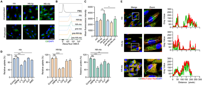
The macropinocytosis, caveolae-, and clathrin-mediated endocytosis are three main types of nanoparticles endocytosis [30]. To investigate whether HA-nio utilize cellular uptake pathways beyond the CD44 route, cells were pre-incubated with various inhibitors targeting different endocytic pathways. These included amiloride (AMD) to block macropinocytosis-mediated endocytosis, filipin (FLP) to block caveolae-mediated endocytosis, chlorpromazine (CPZ) to block clathrin-mediated endocytosis, verapamil (VRPM) to inhibit the OCTN-1 transport protein, and low temperature (4 ℃) to suppress energy-dependent endocytosis. The results indicate that the uptake of HA-lip and HA-nio are both energy-dependent processes, in which HA-lip is mainly transported through clathrin-mediated endocytosis, while HA-nio exhibits multiple endocytic pathways including endocytosis mediated by clathrin and macropinocytosis (Fig. 3D). In addition, blocking the EGT specific transporter OCTN-1 with VRPM had negligible effect on the uptake of HA-nio. This implies that EGT when encapsulated by HA-nio, bypasses the OCTN-1 pathway for cellular uptake, which is expected to overcome the decrease in cellular uptake caused by transporter saturation.
Mitochondria are the main sites where EGT usually selectively accumulates to mitigate cellular oxidative damage and chronic inflammation caused during hypoxia. Therefore, we explored the mitochondria accumulation of different formulations (Fig. 3E). The FITC-EGT and C6-labeled nio, HA-lip, and HA-nio were used to study mitochondrial accumulation ability. The hydrophilic property makes FITC-EGT difficult to cross the cell membrane, resulting in limited co-localization between free FITC-EGT and mitochondria (Fig. S2 in Supporting information). However, various lipid carriers demonstrated good localization with mitochondria, with HA-nio being the most effective (Fig. S3 in Supporting information). HA-CD44 entry pathway and electrostatic interaction between positive HA-nio and negative cell membrane facilitated both the cellular and mitochondrial entrapment of EGT overcoming the restriction of OCTN-1.
Collectively, HA-nio possess excellent capabilities for cellular uptake as well as mitochondrial accumulation, which are crucial for the effective anti-photoaging action of their cargo, EGT.
Excessive UV irradiation would evoke ROS generation, which contributes to skin photoaging via DNA damage, inflammation, oxidative stress, and apoptosis [31]. The evaluation of EGT@HA-nio antioxidative and anti-inflammatory potential in vitro was assessed in irradiation-induced photoaging HDFs (Fig. 4A) [32]. To choose a suitable ultraviolet radiation b (UVB) intensity for cell irradiation, we assessed the cytotoxic effect of various UVB intensities on cells (Fig. S4 in Supporting information). The results show cell viability decreased in a dose-dependent manner and that the cell survival rate was 60% when irradiated with UVB at an intensity of 1000 mJ/cm2, which was selected as a UVB lighting condition for subsequent experiments. To determine whether the model was successfully established, we tested senescence-associated β-galactosidase (SA-β-Gal) positive cells and the senescent cell marker protein p16 (Figs. 4B and C, Fig. S5 in Supporting information). Compared with untreated cells, UVB-irradiated HDFs showed increased expression of blue β-galactosidase and p16 protein, indicating a successfully established cell photoaging model.
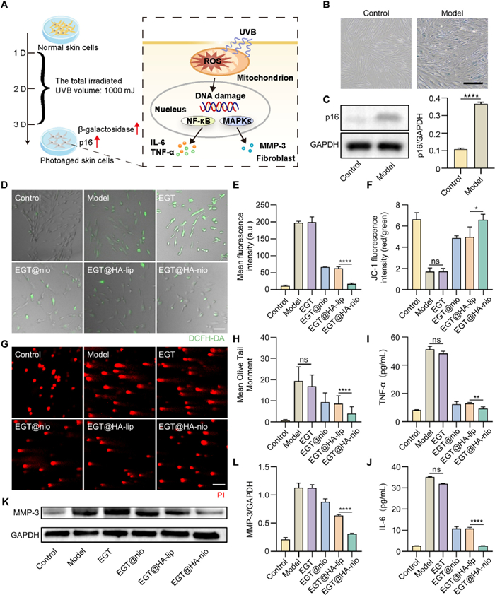
Next, we evaluated the antioxidant capacity of EGT@HA-nio by measuring intracellular ROS levels via the 2′, 7′-dichlorodihydrofluorescein diacetate (DCFH-DA) probe (Figs. 4D and E). As is shown in Fig. 4D, UVB-irradiated cells had the strongest green fluorescence, suggesting that ROS was significantly induced by UVB irradiation. Free EGT can scarcely scavenge ROS due to limited cellular uptake. Compared to EGT@nio or EGT@HA-lip group, the fluorescence intensity of EGT@HA-nio group was much lower, demonstrating that EGT@HA-nio can significantly scavenge ROS.
Since excessive accumulation of ROS may induce mitochondrial dysfunction and DNA damage, the JC-1 probe and comet assay were used to detect mitochondrial transmembrane potential and the situation of DNA strand breakage, respectively. In normal cells with high mitochondrial membrane potential, JC-1 exists in a polymer form and can emit red fluorescence. On the contrary, JC-1 exists in the form of monomers that produce green fluorescence when the mitochondrial membrane potential is low. The ratio of red to green fluorescence represents the proportion of mitochondrial depolarization. Compared to the cell photoaging model, EGT, EGT@nio, or EGT@HA-lip group, the JC-1 fluorescence intensity of the EGT@HA-nio group most closely approximated that of control group, demonstrating that EGT@HA-nio can effectively protect mitochondria from ROS damage, in line with the previous ROS scavenging experiment results (Fig. 4F and Fig. S6 in Supporting information). Similarly, the UVB-irradiated group exhibited obvious comet-like images, indicating that UVB irradiation could cause DNA damage (Fig. 4G). Compared to the photoaging cell model, EGT, EGT@nio, or EGT@HA-lip group, EGT@HA-nio group had less comet-like images but more spherical images, demonstrating that EGT@HA-nio can effectively protect DNA from UV irradiation and ROS damage. The result was also supported by the tail moment parameter (Fig. 4H).
ROS accumulation also activates various signaling pathways, including the nuclear factor kappa-B (NF-κB) and mitogen activated protein kinase (MAPK) pathways, which then promote the expression of inflammatory mediators and ultimately accelerate extracellular matrix (ECM) degradation [33,34]. We analyzed the expression level of interleukin-6 (IL-6) and tumor necrosis factor-α (TNF-α) in HDFs treated with different formulations (Figs. 4I and J). The levels of IL-6 and TNF-α were significantly increased after UVB irradiation and the lowest IL-6 and TNF-α levels were shown in the EGT@HA-nio treated group compared with other treatment groups, suggesting that EGT@HA-nio could inhibit inflammation by reducing the release of proinflammatory cytokines. The activation of matrix metalloproteinases (MMPs) leads to excessive degradation of collagen and elastin in the dermis, resulting in skin aging symptoms such as wrinkles and decreased elasticity. Western blot was performed to detect MMP-3 expression levels in cells (Figs. 4K and L). The results show that EGT@HA-nio treatment induced the most effective downregulation of MMP-3, indicating that it may effectively protect UV-irradiated skin from excessive degradation of collagen and elastin.
In brief, benefiting from more intracellular accumulation of EGT@HA-nio into mitochondria through the HA-CD44 axis and electrostatic interaction, EGT@HA-nio could scavenge ROS to improve mitochondrial dysfunction, reduce DNA damage, and downregulate the expression of IL-6, TNF-α as well as MMP-3 effectively, demonstrating the potential to protect the skin from UV damage.
Encouraged by the enhanced penetration, better cellular uptake, and in vitro ROS scavenging and anti-inflammatory properties, we further evaluated the anti-UV-induced skin damage effect of EGT@HA-nio in the photoaging mice model. BALB/c-nu mice were treated with different methods and irradiated with UV five times a week to establish a photoaging animal experimental model [35]. The treatment process is shown in Fig. 5A. The skin surface of the mice in the control group was smooth, wrinkle-free, and elastic, while the skin of the mice in the model group after long-term UV irradiation was dry, rough, wrinkled, erythematous. The skin damage, roughness, and erythema of the mice in the preparation group were significantly reduced, indicating that the preparation can reduce photoaging damage (Fig. S7 in Supporting information).
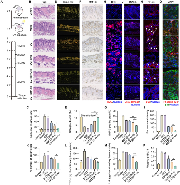
The occurrence of photoaging can lead to skin inflammation and epidermal thickening [36,37]. During this process, collagen degradation increases, destroying the dynamic balance of the ECM due to the upregulation of MMPs expressed in the matrix surrounding collagen [38,39]. The histological profile of the photoaging model was analyzed by hematoxylin and eosin (H&E) staining, Masson's trichrome staining, and Sirius red staining to evaluate the anti-photoaging effect of EGT@HA-nio. H&E staining and epidermal thickness results showed that after UV irradiation in the model group and EGT group, the epidermal thickness increased nearly 5 times, a large number of inflammatory cell infiltrates could be seen in the skin tissue, and the connection structure was relatively loose. The EGT@HA-nio treatment group had less inflammatory cell infiltration and better matrix connection, and there was no significant difference in epidermal thickness between the EGT@HA-nio treatment group and the normal group. This shows that EGT@HA-nio treatment significantly alleviated the skin damage caused by UV irradiation and simultaneously reduced the epidermal thickness (Figs. 5B and C). Masson's trichrome staining method shows that the dermis of normal mouse skin contains abundant collagen fibers, which are blue; the skin of mice in the model group was damaged by UV irradiation, the epidermis fell off, and the collagen fibers were broken and arranged loosely and disorderly. The number of blue-dyed collagen fibers in the skin tissue of mice in the EGT@HA-nio group was significantly improved compared with the control group (Fig. S8 in Supporting information). Sirius red staining further verified the above results and analyzed them quantitatively (Figs. 5D and E). UV irradiation reduced the type Ⅲ collagen area in skin tissue from 10.72% to 3.85%. The type Ⅲ collagen area in the skin tissue of mice in the free EGT treatment group was only 2.52%, while the type Ⅲ collagen area in the treatment group increased significantly and returned to healthy levels. Next, we evaluated the expression of MMP-3 in EGT@HA-nio-treated HDF by immunohistochemical staining (Figs. 5F and G). Compared with the EGT@HA-lip group, EGT@HA-nio significantly down-regulated UV-induced MMP-3 overproduction expression, thereby effectively inhibiting the degradation of collagen. The above experimental results strongly show that EGT@HA-nio effectively mitigated UV-induced skin photoaging and enhanced collagen deposition, indicating HA-nio as a promising platform for transdermal EGT delivery.
To verify the pharmacological mechanism of EGT@HA-nio at the animal level, we assessed its ROS scavenging and anti-inflammatory effects in vivo. Dihydroethidium (DHE) staining results show a significant reduction in ROS levels in the EGT@HA-nio group after UV irradiation (Figs. 5H and I). In vivo experiments further reveal that UV-induced DNA damage led to significant cell apoptosis, as indicated by TdT-mediated dUTP nick-end labeling (TUNEL) immunohistochemistry (Figs. 5J and K). Among the treatment groups, EGT@HA-nio exhibited the strongest protection against UV-induced apoptosis, consistent with comet assay results (Figs. 4G and H). Additionally, enzyme-linked immunosorbent assay (ELISA) results demonstrate that the increased levels of pro-inflammatory cytokines (TNF-α and IL-6), induced by UV irradiation, were reduced by 61.17% and 56.02%, respectively, in the EGT@HA-nio group compared to the model group (Figs. 5L and M). Based on the anti-inflammatory and ROS scavenging effects of the EGT@HA-nio, we further elucidate the mechanism by which the EGT@HA-nio improves UV-induced skin damage. Here we used immunofluorescence staining to study the NF-κB nuclear translocation and the expression of phospho-p38 MAPK/p38 MAPK proteins (Figs. 5N–P, Figs. S9 and S10 in Supporting information). Compared with the normal group, the p65 subunit of NF-κB in the UVB-irradiated skin tissue obviously traveled from the cytosol to the nucleus and the level of phospho-p38 MAPK/p38 MAPK was significantly upregulated. The result indicated that UVB could activate NF-κB and p38 MAPK pathways. Among all treatment groups, the EGT@HA-nio treatment group showed the weakest nuclear fluorescence of NF-κB p65 subunit and the expression of p-p38 MAPK/p38 MAPK protein was the lowest. These findings highlight the superior capacity of EGT@HA-nio to protect skin from UV damage by alleviating oxidative stress and inhibiting inflammation, which is related to NF-κB and MAPK pathway. Overall, EGT@HA-nio can restore the normal level of collagen, maintain the normal thickness and morphology of the epidermis, and have a better anti-photoaging effect than EGT@HA-lip. Notably, deformable HA-nio demonstrate enhanced skin permeability and EGT delivery due to elasticity property provided by inserted S2 molecule and targeting ability conferred by HA modification, thereby improving the delivery of EGT to HDFs and triggering a series of therapeutic effects.
In conclusion, we presented HA-coated niosomes with high deformability to overcome multiple physiological barriers. The single-tail surfactant acted as an edge activator in HA-nio imparted it with excellent elasticity for intact passage through the SC, while HA modification synergistically facilitates targeted intracellular delivery of EGT into mitochondria. This results in effective antioxidative, anti-inflammatory, and anti-photoaging effects against UV-induced skin damage. HA-nio provide a noninvasive approach to across multiple skin barriers and are appropriate for long-term protection compared with other conventional TDDS. For clinical translation, HA-nio with process able to be industrialized and safe raw materials has good application prospects. The superior penetrating performance of HA-nio across multiple skin biological barriers suggests the potential to traverse other critical barriers, including deep tumor tissue, mucus layers, and bacterial biofilms. Therefore, they offer an alternative option for diseases with inadequate drug penetration, such as skin conditions, cancers, and bacterial infections.
The authors declare that they have no known competing financial interests or personal relationships that could have appeared to influence the work reported in this paper.
Lijun Li: Writing – review & editing, Writing – original draft, Visualization, Validation, Methodology, Investigation, Formal analysis, Data curation. Chenliang Guo: Writing – original draft, Validation, Methodology, Formal analysis, Data curation. Yuelin Fang: Writing – review & editing, Writing – original draft, Conceptualization. Zijian Cheng: Writing – original draft, Investigation, Conceptualization. Yaowei Li: Investigation. Zhangyu Wang: Writing – original draft, Investigation. Dian Cai: Writing – original draft, Investigation. Yuqi Xu: Investigation. Wenqi Liu: Writing – original draft. Shouwei Ma: Supervision, Project administration. Xinxin Zhang: Supervision, Resources, Project administration, Funding acquisition, Conceptualization.
The study was supported by the National Natural Science Foundation of China (No. 82222066), and the National Key Research and Development Program of China (No. 2022YFC2304104). We also thank the Electron Microscopy System and the Integrated Laser Microscopy System at the National Facility for Protein Science in Shanghai (NFPS), Zhangjiang Lab, China for providing technical support and assistance in data collection and analysis for cryo-TEM, confocal microscopy, and flow cytometry.
Supplementary material associated with this article can be found, in the online version, at doi:
J. Lee, G.W. Hwang, B.S. Lee, et al., ACS Nano 18 (2024) 5311–5321.
X. Hu, N. Cheng, J. Zhao, et al., Asian J. Pharm. Sci. 14 (2019) 305–312.
K.T.M. Tran, T.D. Gavitt, N.J. Farrell, et al., Nat. Biomed. Eng. 5 (2021) 998–1007.
C.C. Yu, A. Shah, N. Amiri, et al., Adv. Mater. 35 (2023) e2300066. doi: 10.1002/adma.202300066
X. Hu, J. Yu, L. Sun, et al., Chin. Chem. Lett. 35 (2024) 109466. doi: 10.1016/j.cclet.2023.109466
Y. Zhou, D. Gao, Y. Wang, et al., Chin. Chem. Lett. 35 (2024) 108967. doi: 10.1016/j.cclet.2023.108967
Y. Fang, G. Li, C. Huang, et al., Int. J. Biol. Macromol. 229 (2023) 123–135. doi: 10.1016/j.ijbiomac.2022.12.046
Y. Fang, T. Nie, G. Li, et al., Chem. Eng. J. 480 (2024) 147930. doi: 10.1016/j.cej.2023.147930
X. Zhang, M. Zhao, N. Cao, et al., Biomater. Sci. 8 (2020) 1885–1896. doi: 10.1039/c9bm01927h
S. Zhang, P. Xin, Q. Ou, et al., J. Mater. Chem. B 6 (2023) 6723–6730. doi: 10.3233/jifs-221903
C. Wiraja, Y. Zhu, D.C.S. Lio, et al., Nat. Commun. 10 (2019) 1147. doi: 10.1038/s41467-019-09029-9
Z. Lu, S. Du, J. Li, et al., Adv. Mater. 35 (2023) e2303388. doi: 10.1002/adma.202303388
Y. Yang, L. Xu, D. Jiang, et al., Adv. Funct. Mater. 31 (2021) 2104092. doi: 10.1002/adfm.202104092
T.H. Truong, K.P. Alcantara, B.P.I. Bulatao, et al., Carbohydr. Polym. 288 (2022) 119401. doi: 10.1016/j.carbpol.2022.119401
N. Loza-Rodríguez, A. Millán-Sánchez, M. Mallandrich, et al., Pharmaceutics 16 (2024) 1187. doi: 10.3390/pharmaceutics16091187
A. Garcês, M.H. Amaral, J.M. Sousa Lobo, et al., Eur. J. Pharm. Sci. 112 (2018) 159–167. doi: 10.1016/j.ejps.2017.11.023
P. Gurnani, C. Sanchez-Cano, H. Xandri-Monje, et al., Small 18 (2022) 2203070. doi: 10.1002/smll.202203070
X. Zhou, Y. Hao, L. Yuan, et al., Chin. Chem. Lett. 29 (2018) 1713–1724. doi: 10.1016/j.cclet.2018.10.037
S. Polaka, V. Makwana, N. Vasdev, et al., J. Control. Release 345 (2022) 385–404. doi: 10.1016/j.jconrel.2022.03.013
L. Chen, L. Zhang, X. Ye, et al., Protein Cell 15 (2024) 191–206. doi: 10.1093/procel/pwad048
D. Gründemann, S. Harlfinger, S. Golz, et al., Proc. Natl. Acad. Sci. U. S. A. 102 (2005) 5256–5261. doi: 10.1073/pnas.0408624102
S.A.T. Opatha, V. Titapiwatanakun, R. Chutoprapat, Pharmaceutics 12 (2020) 855. doi: 10.3390/pharmaceutics12090855
E. Bárány, M. Lindberg, M. Lodén, Int. J. Pharm. 195 (2000) 189–195. doi: 10.1016/S0378-5173(99)00388-9
S. Ghanbarzadeh, A. Khorrami, S. Arami, Drug Deliv. 22 (2015) 1071–1077. doi: 10.3109/10717544.2013.873837
M.A. Wagdi, A. Salama, M.A. El-Liethy, et al., J. Drug Deliv. Sci. Technol. 84 (2023) 104456. doi: 10.1016/j.jddst.2023.104456
B. Baroli, J. Pharm. Sci. 99 (2010) 21–50. doi: 10.1002/jps.21817
C. Ni, Z. Zhang, Y. Wang, et al., J. Control. Release 357 (2023) 432–443. doi: 10.1016/j.jconrel.2023.03.049
H.M. Liu, W. Tang, X.Y. Wang, et al., Molecules 28 (2023) 1648. doi: 10.3390/molecules28041648
L. Li, C. Liu, J. Fu, et al., Int. J. Biol. Macromol. 243 (2023) 125239. doi: 10.1016/j.ijbiomac.2023.125239
J.J. Rennick, A.P.R. Johnston, R.G. Parton, Nat. Nanotechnol. 16 (2021) 266–276. doi: 10.1038/s41565-021-00858-8
A. Kuzumi, A. Yoshizaki-Ogawa, T. Fukasawa, et al., Am. J. Clin. Dermatol. 25 (2024) 951–966. doi: 10.1007/s40257-024-00891-y
W. Wen, J. Chen, L. Ding, et al., Arch. Biochem. Biophys. 657 (2018) 31–40. doi: 10.1016/j.abb.2018.09.007
L. Li, Y. Wang, Y. Xu, et al., J. Control. Release 371 (2024) 298–312. doi: 10.1016/j.jconrel.2024.05.048
Y. Tian, H. Shi, D. Zhang, et al., J. Control. Release 364 (2023) 618–631. doi: 10.1016/j.jconrel.2023.10.018
Y. You, Y. Tian, Z. Yang, et al., Nat. Biomed. Eng. 7 (2023) 887–900. doi: 10.1038/s41551-022-00989-w
K. Yao, Y. Peng, Q. Tang, et al., Int. J. Nanomed. 19 (2024) 9161–9174. doi: 10.2147/ijn.s446090
H. Zhang, X. Xiao, L. Wang, et al., Sig. Transduc. Target. Ther. 9 (2024) 294. doi: 10.1109/cyberscitech64112.2024.00053
J.S. Choi, W.L. Cho, Y.J. Choi, et al., J. Extracell. Vesicles 8 (2019) 1565885. doi: 10.1080/20013078.2019.1565885
Z. Yan, T. Kavanagh, R. da Silva Harrabi, et al., Adv. Funct. Mater. 34 (2024) 2309711. doi: 10.1002/adfm.202309711

Scheme 1 Schematic illustration of EGT@HA-nio preparation (A) and their therapeutic effect against UV-induced skin damage (B).

Figure 1 Characterization of HA-nio. (A) The composition of HA-nio. (B) The optimal ratio of chol to S2 (phos) in the formulations was analyzed by size and PDI. (C) Representative TEM images of nio, HA-lip, and HA-nio. Scale bar: 100 nm. (D) Size distribution of nio, HA-lip, and HA-nio, as observed via dynamic light scattering. (E) Zeta potential of nio, HA-lip, and HA-nio. (F) Encapsulation efficiency of nio, HA-lip, and HA-nio. (G) Cell viability of HDFs after nio, HA-lip, or HA-nio treatment. (H) Young's modulus (elastic modulus) of nio, HA-lip, and HA-nio was calculated via the Sneddon model. AFM images of nio (I), HA-lip (J), and HA-nio (K) in a liquid environment under different forces. Scale bar: 200 nm. Force–indentation curves of nio (L), HA-lip (M), and HA-nio (N), as observed via AFM. Data are displayed as the mean ± SD (n = 3). ns, no significant. P < 0.05.

Figure 2 Evaluation of the penetration of HA-nio. (A) Schematic illustration of transdermal process. (B) FRET study. (C) Cumulative release curve of EGT of different formulations. (D) Penetration ability of different formulations in BALB/c-nu mice. Scale bar: 500 µm. (E) Penetration ability of HA-nio in pig skin-Franz diffusion cell system. Scale bar: 200 µm. Data are displayed as the mean ± SD (n = 3).

Figure 3 Cellular uptake and mitochondrial accumulation ability of HA-nio. (A) Representative CLSM images of C6-labeled nanovesicles in HDFs. Scale bar: 25 µm. DAPI, 4′, 6-diamidino-2-phenylindole. (B) Representative FCM histograms. (C) Quantitative analysis of FCM. (D) The mechanism of internalization of nio, HA-lip, and HA-nio. (E) Mitochondrial accumulation nio, HA-lip, and HA-nio. Scale bar: 5 µm. Data are displayed as the mean ± SD (n = 3). P < 0.05, **P < 0.01, ***P < 0.001, ****P < 0.0001.

Figure 4 Antioxidative and anti-inflammatory effect of EGT@HA-nio. (A) Establishment of the cell photoaging model. (B) SA-β-Gal staining of UVB irradiated HDFs. Scale bar: 200 µm. (C) The level of p16 protein in the UVB irradiated HDFs was quantified using Western blot. (D) ROS detection in HDFs after the different treatments. Scale bar: 100 µm. (E) Mean fluorescence intensity of ROS production in HDFs after the different treatments. (F) Effect of different treatments on mitochondrial transmembrane potential (ΔΨ). (G) Effect of different treatments on DNA damage through comet assay. Scale bar: 100 µm. (H) Comet parameters in HDFs after the different treatments. Expression of TNF-α (I) and IL-6 (J) after different treatments by ELISA. (K, L) MMP-3/glyceraldehyde-3-phosphate dehydrogenase (GAPDH) expression levels in cells were calculated using Western blot analysis. Data are displayed as the mean ± SD (n = 3 in all quantitative data except for Fig. 4H, n = 50 in Fig. 4H). P < 0.05, **P < 0.01, ****P < 0.0001.

Figure 5 In vivo anti-UV-induced skin damage effect of EGT@HA-nio. (A) Establishment of mice skin photoaging model. (B) H&E staining of skin tissues in different groups. Scale bar: 200 µm. (C) Quantitative analysis of epidermal thickness in different groups. (D) Sirius red staining of skin tissues in different groups. Scale bar: 200 µm. (E) Quantitative analysis of collagen-3 positive areas per field in different groups. (F) MMP-3 immunohistochemistry of skin tissues in different groups. Scale bar: 200 µm. (G) Quantitative analysis of MMP-3 positive areas per field in different groups. (H) DHE staining of skin tissues in different groups. Scale bar: 200 µm. (I) Mean fluorescence intensity of ROS in skin tissues after the different treatments. (J) TUNEL staining of skin tissues in different groups 6 weeks post-irradiation. Scale bar: 200 µm. (K) Quantitative analysis of TUNEL-positive cells. Expression of TNF-α (L) and IL-6 (M) in skin tissue after different treatments. (N) The nuclear translocation of NF-κB subunit p65 in skin tissues after the different treatments. Scale bar: 20 µm. Phospho-p38 MAPK and p38 MAPK immunofluorescence of skin tissues in different groups (O) and quantitative analysis (P). Scale bar: 50 µm. Data are displayed as the mean ± SD (n = 3). P < 0.05, **P < 0.01, ***P < 0.001.

 扫一扫看文章

扫一扫关注我们
 DownLoad:
 下载:
下载:
 下载:
下载: